Granuloma annulare
| Granuloma annulare | |
|---|---|
 | |
| Perforating form of Granuloma annulare on hand | |
| Specialty | Dermatology |
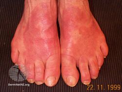
| Symptoms | Clusters of reddish bumps arranged in a circle with a dusky centre and rolled edge, on backs of hands and feet.[1] |
| Usual onset | Children, young adults[1] |
| Causes | Unknown[1] |
| Treatment | Topical corticosteroid[1] |
| Frequency | Females>males[2] |
Granuloma annulare is an inflammation of skin that generally presents as a cluster of small reddish bumps arranged in a circle or ring, typically on the backs of hands and feet.[1] The edge may appear rolled, the centre dusky, and scale is absent.[3]
It has been associated with diabetes and thyroid disease, but the cause is unknown.[1]
Treatment usually involves applying topical corticosteroid.[1]
It is more common in children and young adults.[1] Females are affected more frequently than males.[2]
Signs and symptoms
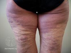
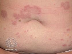
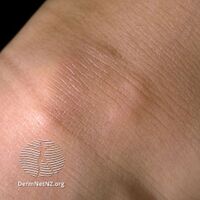
Aside from the visible rash, granuloma annulare is usually asymptomatic. Sometimes the rash may burn or itch. People with granuloma annulare usually notice a ring of small, firm bumps (papules) over the backs of the forearms, hands or feet, often centered on joints or knuckles. The bumps are caused by the clustering of T cells below the skin. These papules start as very small, pimple looking bumps, which spread over time from that size to dime, quarter, half-dollar size and beyond. Occasionally, multiple rings may join into one. Rarely, granuloma annulare may appear as a firm nodule under the skin of the arms or legs. It also occurs on the sides and circumferential at the waist and without therapy can continue to be present for many years. Outbreaks continue to develop at the edges of the aging rings.[citation needed]
-
Granuloma annulare
-
Granuloma annulare
-
Granuloma annulare
-
Granuloma annulare
.jpg)
.jpg)
.jpg)
.jpg)
Causes
The condition is usually seen in otherwise healthy people. Occasionally, it may be associated with diabetes or thyroid disease. It has also been associated with auto-immune diseases such as systemic lupus erythematosus, rheumatoid arthritis, Lyme disease and Addison's disease. At this time, no conclusive connection has been made between patients.[citation needed]
Pathology

Granuloma annulare microscopically consists of dermal epithelioid histiocytes around a central zone of mucin—a so-called palisaded granuloma.[citation needed]
Diagnosis
Types
Granuloma annulare may be divided into the following types:[4]: 703–5
Treatment
Because granuloma annulare is usually asymptomatic and self-limiting with a course of about 2 years, initial treatment is generally topical steroids or calcineurin inhibitors; if unimproved with topical treatments, it may be treated with intradermal injections of steroids. If local treatment fails it may be treated with systemic corticosteroids.[5][6] Treatment success varies widely, with most patients finding only brief success with the above-mentioned treatments. New research out of India suggests that the combination of rifampin (600 mg), ofloxacin (400 mg), and minocycline hydrochloride (100 mg) once monthly, or ROM therapy, produces promising results.[7] Most lesions of granuloma annulare disappear in pre-pubertal patients with no treatment within two years while older patients (50+) have rings for upwards of 20 years. The appearance of new rings years later is not uncommon.[8]
History
The disease was first described in 1895 by Thomas Colcott Fox and it was named granuloma annulare by Henry Radcliffe Crocker in 1902.[9]
See also
References
- ↑ 1.0 1.1 1.2 1.3 1.4 1.5 1.6 1.7 Wakelin, Sarah H. (2020). "22. Dermatology". In Feather, Adam; Randall, David; Waterhouse, Mona (eds.). Kumar and Clark's Clinical Medicine (10th ed.). Elsevier. p. 667. ISBN 978-0-7020-7870-5. Archived from the original on 2021-12-11. Retrieved 2021-12-11.
- ↑ 2.0 2.1 "Granuloma annulare | DermNet". dermnetnz.org. Archived from the original on 3 December 2022. Retrieved 29 March 2023.
- ↑ Dennis, Mark; Bowen, William Talbot; Cho, Lucy (2012). "Granuloma annulare". Mechanisms of Clinical Signs. Elsevier. p. 525. ISBN 978-0729540759. Archived from the original on 2021-07-14. Retrieved 2016-10-18; pbk
{{cite book}}: CS1 maint: postscript (link) - ↑ James, William D.; Berger, Timothy G.; et al. (2006). Andrews' Diseases of the Skin: Clinical Dermatology. Saunders Elsevier. ISBN 0-7216-2921-0.
- ↑ "Archive copy". Archived from the original on 2013-10-17. Retrieved 2013-06-14.
{{cite web}}: CS1 maint: archived copy as title (link) - ↑ "Archive copy". Archived from the original on 2019-08-04. Retrieved 2019-08-04.
{{cite web}}: CS1 maint: archived copy as title (link) - ↑ Marcus, D. V.; Mahmoud, B. H.; Hamzavi, I. H. (2009). "Granuloma annulare treated with rifampin, ofloxacin, and minocycline combination therapy". Archives of Dermatology. 145 (7): 787–9. doi:10.1001/archdermatol.2009.55. PMID 19620560.
- ↑ "Granuloma Annulare: Treatment & Medication - March 14, 2007". Archived from the original on September 16, 2017. Retrieved July 12, 2009.
- ↑ Shanmuga1, Sekar C.; Rai1, Reena; Laila1, A.; Shanthakumari, S.; Sandhya, V. (2010), "Generalized granuloma annulare with tuberculoid granulomas: A rare histopathological variant", Indian Journal of Dermatology, Venereology and Leprology, 76 (1): 73–75, doi:10.4103/0378-6323.58691, PMID 20061743, archived from the original on 4 September 2019, retrieved 23 May 2010
External links
| Classification | |
|---|---|
| External resources |
- Pages with script errors
- CS1 maint: postscript
- CS1 maint: archived copy as title
- All articles with unsourced statements
- Articles with unsourced statements from September 2020
- Articles with invalid date parameter in template
- Monocyte- and macrophage-related cutaneous conditions
- Ailments of unknown cause
- Rare diseases