Dermatofibroma
| Dermatofibroma | |
|---|---|
| Other names: Fibroma durum,[1] dermal dendrocytoma,[2] Fibrous dermatofibroma,[3] Fibrous histiocytoma,[3] Fibroma simplex,[2] Nodular subepidermal fibrosis,[2] and Sclerosing hemangioma[2]) | |
.jpg) | |
| Dermatofibroma | |
| Specialty | Dermatology[4] |
| Symptoms | Small bump in the skin[4] |
| Duration | Long term[4] |
| Causes | Trauma[4] |
| Risk factors | Lupus, HIV, blood cancer, medicines that weaken immunity.[4] |
| Diagnostic method | Direct vision, rarely biopsy[4] |
| Differential diagnosis | Granular cell tumor, melanoma, clear cell acanthoma, dermatofibrosis lenticularis disseminata[4] |
| Treatment | Usually none[4] |
A dermatofibroma, or benign fibrous histiocytomas, is a type of non-cancerous soft tissue tumor that appears as a bump in the skin.[1] It is typically found on the legs, elbows and chest of an adult.[4] It does not usually hurt.[4]
Its size usually ranges from 0.2cm to 2cm, and have been reported to be larger.[4] It typically results from mild trauma such as an insect bite.[4] Risk factors for developing multiple dermatofibromas include lupus, HIV, blood cancer and some medicines that weaken immunity.[4]
It is usually diagnosed by its appearance, but a biopsy may be required.[4] Other bumps such as granular cell tumor, melanoma, clear cell acanthoma and dermatofibrosis lenticularis disseminata may look similar.[4] Reassurance is generally given and usually no treatment is needed.[4] It can remain unchanged for years, but can resolve spontaneously.[4]
Signs and symptoms
Dermatofibromas[5] are hard solitary slow-growing papules (rounded bumps) that may appear in a variety of colours, usually brownish to tan; they are often elevated or pedunculated. A dermatofibroma is associated with the dimple sign; by applying lateral pressure, there is a central depression of the dermatofibroma. Although typical dermatofibromas cause little or no discomfort, itching and tenderness can occur. Dermatofibromas can be found anywhere on the body, but most often they are found on the legs and arms.[6] They occur most often in women; the male to female ratio is about 1:4.[7] The age group in which they most commonly occur is 20 to 45 years.
Some physicians and researchers believe dermatofibromas form as a reaction to previous injuries such as insect bites or thorn pricks.[7] They are composed of disordered collagen laid down by fibroblasts. Dermatofibromas are classed as benign skin lesions, meaning they are completely harmless, though they may be confused with a variety of subcutaneous tumours.[8] Deep penetrating dermatofibromas may be difficult to distinguish, even histologically, from rare malignant fibrohistocytic tumours like dermatofibrosarcoma protuberans.[9]
Dermatofibromas typically have a positive buttonhole sign, or central dimpling in the center.[10]
-
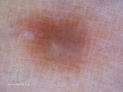
Dermatoscopy of dermatofibroma
-
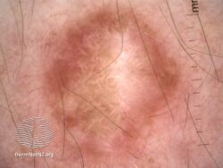
Dermatofibroma dermatoscopy
-
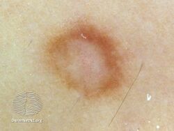
Dermatoscopy of dermatofibroma
.jpg)
.jpg)
.jpg)
Diagnosis
Immunohistochemical staining

| Neoplasm | CD34[2] | Stromelysin-3[11] | Factor XIIIa[7] |
|---|---|---|---|
| Dermatofibroma | - | + | + |
| Dermatofibrosarcoma protuberans | + | - | - |
References
- ↑ 1.0 1.1 DE, Elder; D, Massi; RA, Scolyer; R, Willemze (2018). "5. Soft tissue tomours: Dermatofibroma". WHO Classification of Skin Tumours. Vol. 11 (4th ed.). Lyon (France): World Health Organization. pp. 310–312. ISBN 978-92-832-2440-2. Archived from the original on 2022-07-11. Retrieved 2022-09-06.
- ↑ 2.0 2.1 2.2 2.3 2.4 Rapini, Ronald P.; Bolognia, Jean L.; Jorizzo, Joseph L. (2007). Dermatology: 2-Volume Set. St. Louis: Mosby. ISBN 978-1-4160-2999-1.[page needed]
- ↑ 3.0 3.1 Freedberg; et al. (2003). Fitzpatrick's Dermatology in General Medicine (6th ed.). McGraw-Hill. p. 668. ISBN 978-0-07-138076-8.
- ↑ 4.00 4.01 4.02 4.03 4.04 4.05 4.06 4.07 4.08 4.09 4.10 4.11 4.12 4.13 4.14 4.15 4.16 James, William D.; Elston, Dirk; Treat, James R.; Rosenbach, Misha A.; Neuhaus, Isaac (2020). "28. Dermal and subcutaneous tumors". Andrews' Diseases of the Skin: Clinical Dermatology (13th ed.). Edinburgh: Elsevier. pp. 617–618. ISBN 978-0-323-54753-6. Archived from the original on 2022-03-01. Retrieved 2022-03-01.
- ↑ "Dermatofibroma | Genetic and Rare Diseases Information Center (GARD) – an NCATS Program". rarediseases.info.nih.gov. Archived from the original on 2020-10-17. Retrieved 2018-04-17.
- ↑ "dermatofibroma" at Dorland's Medical Dictionary
- ↑ 7.0 7.1 7.2 Dermatofibroma at eMedicine
- ↑ Jung, Kyu Dong; Lee, Dong-Youn; Lee, Joo-Heung; Yang, Jun-Mo; Lee, Eil-Soo (2011). "Subcutaneous Dermatofibroma". Annals of Dermatology. 23 (2): 254–7. doi:10.5021/ad.2011.23.2.254. PMC 3130878. PMID 21747634.
- ↑ Hanly, A. J.; Jordà, M; Elgart, G. W.; Badiavas, E; Nassiri, M; Nadji, M (2006). "High proliferative activity excludes dermatofibroma: Report of the utility of MIB-1 in the differential diagnosis of selected fibrohistiocytic tumors". Archives of Pathology & Laboratory Medicine. 130 (6): 831–4. doi:10.1043/1543-2165(2006)130[831:HPAEDR]2.0.CO;2 (inactive 2021-01-10). PMID 16740036.
{{cite journal}}: CS1 maint: DOI inactive as of January 2021 (link) - ↑ Boursicot, Katharine (24 January 2013). Oxford Assess and Progress: Clinical Specialties. Oxford University Press. p. 249. ISBN 9780199657582.
- ↑ Kim, H.J.; Lee, J.Y.; Kim, S.H.; Seo, Y.J.; Lee, J.H.; Park, J.K.; Kim, M.H.; Cinn, Y.W.; Cho, K.H.; Yoon, T.Y. (2007). "Stromelysin-3 expression in the differential diagnosis of dermatofibroma and dermatofibrosarcoma protuberans: Comparison with factor XIIIa and CD34". British Journal of Dermatology. 157 (2): 319–24. doi:10.1111/j.1365-2133.2007.08033.x. PMID 17596171. S2CID 7049937.
External links
| Classification | |
|---|---|
| External resources |