Epidermoid cyst
| Epidermoid cyst | |
|---|---|
| Other names: Epidermal cyst, epidermal inclusion cyst, keratin cyst, infundibular cyst[1] | |
.jpg) | |
| Epidermoid cyst | |
| Specialty | Dermatology |
| Symptoms | Painless, unless ruptured or infected, central blackhead[2] |
| Complications | Cancer[3] |
| Usual onset | 30s and 40s[3] |
| Duration | Long-term[3] |
| Causes | Random, Gardner syndrome, Gorlin syndrome, cyclosporine[3] |
| Diagnostic method | Based on symptoms and examination[1] |
| Differential diagnosis | Lipoma, dermoid cyst, acne, trichilemmal cyst, milia[1][3] |
| Treatment | None, incision and drainage, surgical removal[1] |
| Frequency | Relatively common[3] |
Epidermoid cysts are generally non-serious skin cysts.[3] They typically do not result in pain, unless an abscess forms or they rupture internally.[2] A dark pore may be present.[3] They are most common on the face, neck, and trunk; though may form anywhere.[3] They often gradually grow over years.[3] Rare complications may include cancer.[3]
Generally they occur randomly; though are associated with Gardner syndrome, Gorlin syndrome, and the medication cyclosporine.[3] They occur under the epidermis and are filled with keratin.[3] Diagnosis is generally based on symptoms and examination.[1]
Specific treatment is not required in those without symptoms.[1] Larger cysts may be removed surgically, with efforts to also remove the capsule.[1] If infected, incision and drainage together with antibiotics may be indicated.[1]
They are the most common type of cyst.[3] Most often they occur in peoples 30s and 40s.[3] They effect males twice as often as females.[3] They have incorrectly been referred to as a "sebaceous cyst", as they do not contain sebum.[1]
Signs and symptoms
The epidermoid cyst may have no symptoms, or it may be painful when touched. It can release macerated keratin (cheesy material). In contrast to pilar cysts, epidermoid cysts are usually present on parts of the body with relatively little hair.[4] An epidermoid cyst can occur as a vaginal cysts.[5]
Although they are not malignant, there are rare cases of malignant tumors arising from an epidermoid cyst.[6] Epidermal inclusion cysts account for approximately 85–95% of all excised cysts, malignant transformation is exceedingly rare. The incidence of squamous cell carcinoma developing from an epidermal inclusion cyst has been estimated to range from 0.011 to 0.045%.[7]
-
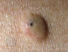
Epidermal cyst with a central poor
-

Epidermal cyst on the neck, inflamed
-
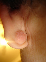
Epidermal cyst in the earlobe
-
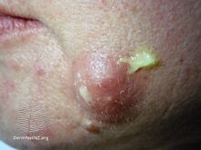
Discharge from the cyst with palpation
.jpg)


.jpg)
Causes

Epidermoid cysts commonly result from implantation of epidermis into the dermis, as in trauma or surgery. They can also be caused by a blocked pore adjacent to a body piercing. They are also seen in Gardner's syndrome and Nevoid basal-cell carcinoma syndrome on the head and neck. They can be infected by bacteria and form a pimple-like shape.
Diagnosis
Epidermoid cysts are usually diagnosed when a person notices a bump on their skin and seeks medical attention. The definitive diagnosis is made after excision by a pathologist based on microscopic appearance of a cystic lesion lined by cornified epithelium containing lamellated keratin without calcifications. They can also be seen as isointense lesions on MRI or hyperintensities on FLAIR.
-

CT scan, showing a homogenous hypodense volume (unspecific cyst-like)
-

Epidermoid cyst in a testicle on ultrasound, with lamellated ("onion skin") appearance
-
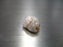
Macroscopic appearance of a resected (surgically removed) intracranial cyst, with pearl appearance
-

Histopathology, showing a keratinizing stratified squamous epithelium, and a lumen containing keratin flakes
-

Histopathology showing epithelium and lamellated keratin (left)


Treatment
Cysts can be removed by excision.[8]
In case of fronto-ethmoidal epidermoid cysts, surgical resection appears to be the mainstay of treatment; however, the extent of resection is dictated by adherence of the tumor capsule to the surrounding vital structures.[9]
Hydrogen peroxide gel (H2O2) was previously recommended for cyst treatment, particularly those on body piercings. However the gel cannot adequately permeate the cyst and was not found to be effective.[10] Hydrogen peroxide is no longer recommended for wound care by doctors as it can damage the healing tissues.[11]
On body piercings, self treatment with a hot saline soak to help drain the cyst and the use of an antibacterial or medicated talcum powder to help dry out the bump and reduce bacterial proliferation is generally recommended until medical advice can be obtained.[12] (Use of talc is no longer recommended due to recently discovered associations with multiple cancers.)[citation needed] Piercings, however, are more likely to be victims of hypertrophic scarring than a cyst. Cheek piercings seem to be the piercing most prone to cysts due to the possible interruption of saliva ducts.
-

Surgery of a suprasternal epidermoid cyst, showing a smooth surface
-
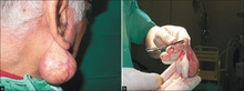
a) Giant earlobe epidermoid cyst b) cyst resection and content


Terminology
Several synonyms exist for epidermoid cysts, including epidermal cyst, infundibular cyst, keratin cyst and epidermal inclusion cyst[13][14]: 778 [15] Epidermal inclusion cyst more specifically refers to implantation of epidermal elements into the dermis. The term infundibular cyst refers to the site of origin of the cyst: the infundibular portion of the hair follicle. The majority of epidermal inclusion cysts originate from the infundibular portion of the hair follicle, thus explaining the interchangeable,[16] yet inaccurate, use of these two terms.
Epidermoid cyst may be classified as a sebaceous cyst,[17] although technically speaking it is not sebaceous.[18] "True" sebaceous cysts, cysts which originate from sebaceous glands and which contain sebum, are relatively rare and are known as steatocystoma simplex or, if multiple, as steatocystoma multiplex. Medical professionals have suggested that the term sebaceous cyst be avoided since it can be misleading.[19]: 31 In practice, however, the term is still often used for epidermoid and pilar cysts.
See also
- Intracranial epidermoid cyst
- List of cutaneous neoplasms associated with systemic syndromes
- Proliferating epidermoid cyst
- Verrucous cyst
References
- ↑ 1.0 1.1 1.2 1.3 1.4 1.5 1.6 1.7 1.8 "Epidermoid cyst | DermNet NZ". dermnetnz.org. Archived from the original on 18 August 2022. Retrieved 11 August 2022.
- ↑ 2.0 2.1 "Cutaneous Cysts - Dermatologic Disorders". Merck Manuals Professional Edition. Archived from the original on 8 June 2022. Retrieved 11 August 2022.
- ↑ 3.00 3.01 3.02 3.03 3.04 3.05 3.06 3.07 3.08 3.09 3.10 3.11 3.12 3.13 3.14 3.15 Zito, PM; Scharf, R (January 2022). "Epidermoid Cyst". PMID 29763149.
{{cite journal}}: Cite journal requires|journal=(help) - ↑ "cysts - British Association of Dermatologists". Archived from the original on 2008-01-10. Retrieved 2007-11-14.
- ↑ Zimmern, Francois Haab, Christopher R. Chapple (2006). Vaginal Surgery for Incontinence and Prolapse. Springer Science & Business Media. p. 271. ISBN 1852339128. Archived from the original on May 9, 2022. Retrieved March 2, 2018.
{{cite book}}: CS1 maint: uses authors parameter (link) - ↑ Jehle KS, Shakir AJ, Sayegh ME (2007). "Squamous cell carcinoma arising in an epidermoid cyst". British Journal of Hospital Medicine. 68 (8): 446. doi:10.12968/hmed.2007.68.8.24499. PMID 17847698.
- ↑ Frank, Ethan; Macias, David; Hondorp, Brian; Kerstetter, Justin; Inman, Jared C. (2018). "Incidental Squamous Cell Carcinoma in an Epidermal Inclusion Cyst: A Case Report and Review of the Literature". Case Reports in Dermatology. 10 (1): 61–68. doi:10.1159/000487794. ISSN 1662-6567. PMID 29681810. Archived from the original on 2020-11-01. Retrieved 2022-01-19.
- ↑ "Minimal excision technique for removal of an epidermoid cyst". Am Fam Physician. 65 (7): 1423–4. 2002. PMID 11996427. Archived from the original on 2008-07-06. Retrieved 2007-11-15.
- ↑ "Epidermoid Cysts in the Frontal Lobe – A Case Series - Journal of Pioneering Medical Sciences". www.jpmsonline.com. Archived from the original on 27 July 2014. Retrieved 2 April 2018.
- ↑ Graziani, F; Vano, M; Tartaro, G; Fanelli, G; Gabriele, M (2003). "The use of hydrogen peroxide in the experimental therapy of cysts. An in vitro analysis". Minerva Stomatologica. 52 (7–8): 373–7, 378–80. PMID 14608258.
- ↑ Walsh, L. J. (2000). "Safety issues relating to the use of hydrogen peroxide in dentistry". Australian Dental Journal. 45 (4): 257–69, quiz 289. doi:10.1111/j.1834-7819.2000.tb00261.x. PMID 11225528.
- ↑ "Archived copy". Archived from the original on 2014-04-23. Retrieved 2014-04-21.
{{cite web}}: CS1 maint: archived copy as title (link) - ↑ Melton, Jason R. Swanson and Jeffrey L. "Epidermoid cyst". www.meddean.luc.edu. Archived from the original on 2020-10-29. Retrieved 2022-01-19.
- ↑ Freedberg, et al. (2003). Fitzpatrick's Dermatology in General Medicine. (6th ed.). McGraw-Hill. ISBN 0-07-138076-0.
- ↑ James, William; Berger, Timothy; Elston, Dirk (2005). Andrews' Diseases of the Skin: Clinical Dermatology. (10th ed.). Saunders. ISBN 0-7216-2921-0.
- ↑ "Epidermoid cyst". Archived from the original on 2007-11-21. Retrieved 2007-11-14.
- ↑ "Epidermoid and pilar cysts (previously known as sebaceous cysts)". British Association of Dermatologists. Archived from the original on February 5, 2016. Retrieved April 2, 2014.
- ↑ "Epidermoid and Pilar Cysts (Sebaceous Cysts) - Patient UK". Archived from the original on 2013-07-06. Retrieved 2013-03-04.
- ↑ Neville BW, Damm DD, Allen CA, Bouquot JE (2002). Oral & maxillofacial pathology (2nd ed.). Philadelphia: W.B. Saunders. ISBN 978-0721690032.
External links
| Classification | |
|---|---|
| External resources |
- Pages with script errors
- CS1 errors: missing periodical
- CS1 maint: uses authors parameter
- CS1 maint: archived copy as title
- All articles with unsourced statements
- Articles with unsourced statements from May 2017
- Articles with invalid date parameter in template
- Epidermal nevi, neoplasms, and cysts
- Conditions of the skin appendages
- RTT