Pilomatricoma
| Pilomatricoma | |
|---|---|
| Other names: Calcifying epithelioma of Malherbe,[1] Malherbe calcifying epithelioma, and Pilomatrixoma | |
.jpg) | |
Pilomatricoma, is a benign skin tumor derived from the hair matrix.[2][3] These neoplasms are relatively uncommon and typically occur on the scalp, face, and upper extremities. Clinically, pilomatricomas present as a subcutaneous nodule or cyst with unremarkable overlying epidermis that can range in size from 0.5-3.0 cm, but the largest reported case was 24 cm.[4]
Signs and symptoms
Pilomatricoma is a small lump underneath the human skin which grows slow and has no symptoms other than the aforementioned.[5]
-
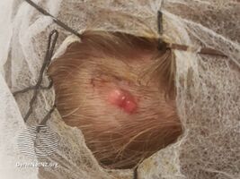
Dermoscopy of pilomatricoma
-
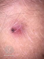
Pilomatricoma dermoscopy
-

Micrograph of a pilomatricoma showing the characteristic "ghost" cells (anucleate squamous cells), benign viable squamous cells and multi-nucleated giant cells. H&E stain.
.jpg)
.jpg)

Associations
Pilomatricomas have been observed in a variety of genetic disorders including Turner syndrome, myotonic dystrophy, Rubinstein-Taybi syndrome, Trisomy 9, and Gardner syndrome.[6] It has been reported that the prevalence of pilomatricomas in Turner syndrome is 2.6%.[7]
Hybrid cysts that are composed of epidermal inclusion cysts and pilomatricoma-like changes have been repeatedly observed in Gardner syndrome.[8][9][10][11] This association has prognostic import, since cutaneous findings in children with Gardner Syndrome generally precede colonic polyposis.[citation needed]
Histologic features
Pilomatricomas consist of anucleate squamous cells (called "ghost cells"), benign viable squamous cells and foreign body giant cells. These neoplasms have a characteristic transition of cells. The lining of the cyst consists of basoloid cells with indistinct cell borders and basophilic nuclei that mature into the eosinophilic anucleated squamous cells. The presence of calcifications with foreign body giant cells is common within the tumors.[12]
Pathogenesis
Pilomatricoma is associated with high levels of beta-catenin caused by either a mutation in the APC gene or a stabilizing mutation in the beta-catenin gene, CTNNB1. High levels of beta-catenin increases cell proliferation, inhibit cell death, and ultimately leads to neoplastic growth.[7]
Diagnosis
This section is empty. You can help by adding to it. (May 2018) |
See also
- Malignant pilomatricoma
- List of cutaneous conditions
- List of cutaneous neoplasms associated with systemic syndromes
References
- ↑ Rapini, Ronald P.; Bolognia, Jean L.; Jorizzo, Joseph L. (2007). Dermatology: 2-Volume Set. St. Louis: Mosby. ISBN 978-1-4160-2999-1.[page needed]
- ↑ James, William Daniel; Berger, Timothy G.; Elston, Dirk M., eds. (2006). Andrews' Diseases of the Skin: Clinical Dermatology. Saunders Elsevier. p. 670. ISBN 978-0-8089-2351-0.
- ↑ Levy, Jaime; Ilsar, Michael; Deckel, Yael; Maly, Alexander; Anteby, Irene; Pe'er, Jacob (2008). "Eyelid Pilomatrixoma: A Description of 16 cases and a Review of the Literature". Survey of Ophthalmology. 53 (5): 526–35. doi:10.1016/j.survophthal.2008.06.007. PMID 18929763.
- ↑ Gongidi, P.; Meshekow, J.; Holdbrook, T.; Germaine, P. (2015). "Giant Pilomatrixoma Presenting in the Posterior Thorax, a Rare Location and the Largest Described". Case Reports in Radiology. 2015: 590742. doi:10.1155/2015/590742. PMC 4339831. PMID 25763287.
- ↑ "Pilomatricoma: MedlinePlus Genetics". medlineplus.gov. Archived from the original on 24 March 2021. Retrieved 19 July 2021.
- ↑ Cooper, Philip H.; Fechner, Robert E. (1983). "Pilomatricoma-like changes in the epidermal cysts of Gardner's syndrome". Journal of the American Academy of Dermatology. 8 (5): 639–44. doi:10.1016/S0190-9622(83)70071-X. PMID 6863619.
- ↑ 7.0 7.1 Glanz, Steven M.; Kessler, Harvey P.; Eskin, Thomas A.; Liu, Chen; Hassanein, Ashraf M. (2003). "b-Catenin Is Expressed Aberrantly in Tumors Expressing Shadow Cells Pilomatricoma, Craniopharyngioma, and Calcifying Odontogenic Cyst". American Journal of Clinical Pathology. 120 (5): 732–6. doi:10.1309/EALEG7LD6W7167PX. PMID 14608900.
- ↑ Narisawa, Yutaka; Kohda, Hiromu (1989). "An Unusual Hybrid Cyst in Gardner's Syndrome with Partial Differentiation toward the Inner Root Sheath". The Journal of Dermatology. 16 (6): 492–5. doi:10.1111/j.1346-8138.1989.tb01591.x. PMID 2628457.
- ↑ Rütten, A; Wenzel, P; Goos, M (1990). "Gardner-Syndrom mit pilomatrixomartigen Haarfollikelzysten" [Gardner syndrome with pilomatrixoma-like hair follicle cysts]. Der Hautarzt; Zeitschrift FüR Dermatologie, Venerologie, und Verwandte Gebiete (in Deutsch). 41 (6): 326–8. PMID 2380070. INIST:19291018.
- ↑ Narisawa, Yutaka; Kohda, Hiromu (1995). "Cutaneous cysts of Gardner's syndrome are similar to follicular stem cells". Journal of Cutaneous Pathology. 22 (2): 115–21. doi:10.1111/j.1600-0560.1995.tb01392.x. PMID 7560342.
- ↑ Urabe, Kazunori; Xia, Jianxin; Masuda, Teiichi; Moroi, Yoichi; Furue, Masutaka; Matsumoto, Takayuki (2004). "Pilomatricoma-Like Changes in the Epidermoid Cysts of Gardner Syndrome with an APC Gene Mutation". The Journal of Dermatology. 31 (3): 255–7. doi:10.1111/j.1346-8138.2004.tb00669.x. PMID 15187352.
- ↑ Elder, David E.; Johnson, Bernett L.; Elenitsas, Rosalie, eds. (2005). Lever's Histopathology of the Skin. Lippincott Williams & Wilkins. ISBN 978-0-7817-3742-5.[page needed]
External links
| Classification | |
|---|---|
| External resources |
- Pages with script errors
- Wikipedia articles needing page number citations from July 2016
- Articles with invalid date parameter in template
- CS1 Deutsch-language sources (de)
- All articles with unsourced statements
- Articles with unsourced statements from July 2016
- Articles to be expanded from May 2018
- All articles to be expanded
- Articles with empty sections from May 2018
- All articles with empty sections
- Articles using small message boxes
- Cutaneous congenital anomalies