Talk:Fallopian tube/Archive 1
| This page is an archive of past discussions. Do not edit the contents of this page. If you wish to start a new discussion or revive an old one, please do so on the current talk page. |
Homology
The list of homologues of the human reproductive system gives appendix testis as a homologue of the fallopian tube, but this article claims there is no known male homologue. Which is true? Leedar 13:29, 27 November 2006 (UTC)
Obvious falsehood
Someone put the following comment on the page without citation... not that it's needed as it's obviously incorrect. I removed it.
"The anatomy of this complex structure was first discovered by Latin-American Gynaecologist Dr. Fallopian E. Tube, and due to his success was awarded the prestigeous title of Gynaecologist of the Year in 1985."
71.234.109.192 00:39, 9 September 2007 (UTC) rhetoric
Afraid to start tampering with the article, as I am not experienced with editing Wikipedia articles. It would be nice if someone more experienced could add in the epithelium that there are three, not two types of cells found there. The third being Basal (stem) cells - undifferentiated cells responsible for regeneration of the epithelium. — Preceding unsigned comment added by 89.73.254.101 (talk) 13:16, 25 September 2011 (UTC)
File:Fallopian tube anatomy.jpg Nominated for speedy Deletion

|
An image used in this article, File:Fallopian tube anatomy.jpg, has been nominated for speedy deletion for the following reason: All Wikipedia files with unknown copyright status
Don't panic; you should have time to contest the deletion (although please review deletion guidelines before doing so). The best way to contest this form of deletion is by posting on the image talk page.
This notification is provided by a Bot --CommonsNotificationBot (talk) 00:15, 7 November 2011 (UTC) |
Oviducts
If oviducts are the same as fallopian tubes, why do they have separate pages?--Jcvamp 16:43, 2 November 2006 (UTC)
I think the pages should be merged.--Jcvamp 14:25, 1 January 2007 (UTC)
- I also think they should be merged. I added a merge template to each. Cmcnicoll (talk) 06:39, 14 February 2010 (UTC)
(just FYI) I think, if you had to merge them though, you would have to specify that oviducts only exist in non-mammalian vertebrates User:4th Opinion: 3:55, 12 April 2011 —Preceding unsigned comment added by 98.180.55.233 (talk)
Taking Biology currently, my professor acknowledges the term fallopian tubes but the correct scientific term is oviduct. For human anatomy at least, the articles should be combined. 24.68.45.183 (talk) 01:35, 2 June 2011 (UTC)
Anthropocentrism in Wikipedia at its worst
- A stupid stupid idea -- oviducts subsume fallopian tubes as a topic, so if anything fallopian tubes should be merged into the oviduct article. However, speaking as a human and a mammal, I think it's nice to have articles on the specific human versions of things.
- From the oviduct article: "In mammals, the portion of the oviduct above the uterus is referred to as the fallopian tube."
- To repeat -- oviducts are NOT the same as fallopian tubes... fallopian tubes are a part of the oviduct in mammals. 75.62.132.92 (talk) 15:20, 6 February 2012 (UTC)
- The two are definitely not the same thing. Any objections to removing the merge template? - Manfi (talk) 12:25, 27 May 2012 (UTC)
Add better schematic picture about the general structure where you can see for instance Isthmus clearly
Example of such picture: https://dl.dropbox.com/u/62073194/Screen%20Shot%202012-06-03%20at%2012.44.48%20PM.png I cannot add it myself because I do not know the source.
- SaminTietokirja, 3. June 2012 — Preceding unsigned comment added by SaminTietokirja (talk • contribs) 09:47, 3 June 2012 (UTC)
Caps?
Why is this capitalized? In other articles as well. -SV|t 10:13, 10 July 2005 (UTC)
- Probably because they are named after someone. Sort of similar to how the ampulla of Vater and the loop of Henle always have the names capitalized. Alex.tan 12:42, July 10, 2005 (UTC)
- "Probably" doesn't cut it. It needs to be in the article, and Id like to see examples of where fallopian is always capitalized. It appears from my readings this is not the case, and even if it is named for someone, its more common that the thing is uncapitalized, while the persons Name is. -SV|t 18:03, 10 July 2005 (UTC)
- Probably will have to cut it until you provide better evidence than just giving your opinion. I did look up dictionary.com and came up with this. I think it's fair enough to keep them capitalised - it's common enough usage as uncapitalised but we should credit who it's named after. For example, Purkinje cells are, AFAIK always capitalised. Alex.tan 00:05, 13 January 2006 (UTC)
I've added a bit on lowercase fallopian. I ought to say that Clinically Oriented Anatomy by Keith L. Moore is probably the textbook for undergrad English-language academic anatomists, and it uses lowercase 'fallopian' exclusively, though it prefers the term 'uterine tube'. As I say in the article, the reason why it's now lowercase is because to many lay people, there is simply no other term for it. - Richardcavell 14:28, 26 March 2006 (UTC)
- The solution to this is fairly simple: use the international anatomical standard (Terminologia Anatomica). The reason why Moore prefers 'uterine tube' is because this is the standard name for it. Well, technically it's 'tuba uterina', but the standard English is uterine tube. In fact, the term 'fallopian tube' has all but disappeared from modern texts, and, like other eponyms, is falling out of use (especially among the non-old). You can check out the Wikipedia Anatomy project.--Mauvila 08:43, 12 July 2006 (UTC)
- They're named after the anatomist and physician Gabriel Fallopius (1523–1562). However, Webster's Dictionary gives it as "fallopian tube", uncapitalised. Take your pick. — QuicksilverT @ 22:38, 7 August 2014 (UTC)
- Have changed to lowercase so this is not focused on in future taking attention away from article content.Charlotte135 (talk) 21:32, 29 November 2015 (UTC)
tunica muscularis
The longitudinal and circular muscular layers are located beneath the subserosa, they are not part of it. Pagurus82 (talk) 07:00, 19 October 2018 (UTC)
Looking
Looking at the embryological proces; shouldn't it say that the tubes go from uterus to ovaria, instead of from ovary to uterus?? I don't think the trvelpath of the oocyt should be the lead on this matter. (ST)
Do the fallopian tubes develop from the same tissue that gives rise to the sperm ducts in males? AxelBoldt 23:33 Jan 2, 2003 (UTC)
No. The Fallopian tubes are Muellerian, the vas deferens Wolffian. -phma
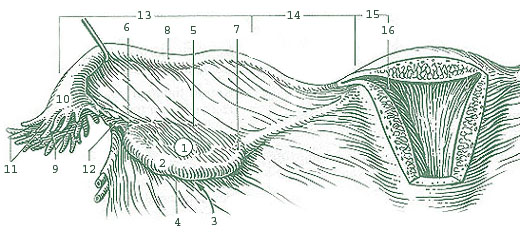
We need a better picture here; the given one does not explain at all where the tubes run and where they attach. AxelBoldt 10:36, 21 Jul 2004 (UTC)
Conflicting information?
Hi. I was reading the Ovulation article before I came to this one, and there I was led to believe that all follicles within the ovaries are called "ovarian follicle", and monthly only one develops into a Graafian follicle, which is what actually releases the ovum into the Fallopian tube when it ruptures (hence the "ovarian follicle" doesn't rupture, but rather the Graafian follicle does). Did I misunderstand something or is there conflicting information between these two articles? On the other hand, the Ovarian follicle article makes no mention at all to the transformation of an ovarian follicle into a Graafian follicle, but the Ovulation article makes it look like a big deal, so at the very least that article is somewhat misleading — or the other two articles are incomplete. Mine is a layman's perspective, which indicates that although the ensemble of the articles may be fine for someone who actually knows that stuff, it doesn't work that well for those who try to start from scratch, such as myself. Regards, Redux 18:56, 4 Oct 2004 (UTC)
Hello. The problem is that the terminology is not used 100% consistently. "Graafian follicle" is an older eponym whose use seems to be falling out of favor. The trend seems to be to refer to all follicles in the ovary as "ovarian follicles" and then to refer to the various stages of development as "primordial follicle," "primary follicle," "vesicular follicle" and "mature follicle," with the last corresponding to the "Graafian follicle." Osmodiar 07:37, 20 Nov 2004 (UTC)
I see. Thanks for the explanation. But don't you think the text of the articles I mentioned should be (slightly) altered to better reflect what you have said. Right now, the layman reader is quite likely to misunderstand that particular aspect of the nomenclature. Regards, Redux 20:46, 10 Dec 2004 (UTC)
peritoneal cavity
I changed "...open into peritoneum" to "...open into peritoneal cavity" because "peritoneum" refers to the layer of tissue derived from mesoderm, while "peritoneal cavity" refers to the actual space in the abdomen. Osmodiar 06:59, 20 Nov 2004 (UTC)
Proposed merge of Ampulla of Fallopian tube into Fallopian tube
The following discussion is closed. Please do not modify it. Subsequent comments should be made on the appropriate discussion page. No further edits should be made to this discussion.
It is not helpful to have these tiny subarticles separate from the main article. It is easier for readers to have them colocated on the main article, and will likely also mean they receive more editing attention. Tom (LT) (talk) 23:36, 28 December 2019 (UTC)
Proposed merge of Ostium/Infundibulum/Fimbriae of uterine tube into Fallopian tube
As above. Additionally, the notability of this being an ostium is entirely derived from its parent structure. Would benefit from merging. Tom (LT) (talk) 23:37, 28 December 2019 (UTC)
- I have professional expertise in Anatomy and concur with your suggestion. I notified the apparent author User:Mikael Häggström/User:Arcadian as a courtesy. I'm not sure how an automatic merge would work with respect to the figure in each subarticle -- it's the same one repeated with different captions. If merging is done manually, I would ask that you definitely preserve the figure from the subarticles -- it's much better than the one in the main article, and apparently better resolution too. I'm not sure that preserving the Identifiers is necessary, but if one is trying to be encyclopedic then they should be preserved. Further, the main article already lists these 4 regions, but it would be a significant loss if the details currently in these subarticles are buried in a mere list (e.g., fimbriae are cilliated, ostium is where most fertilization takes place, etc.). Merging them as subsections may be unnecessary but it might be better for them simply to appear as separate, discrete paragraphs.Lapabc (talk) 23:13, 26 February 2020 (UTC)
- BTW, since you're monitoring this article Tom (LT), I believe the banner that it does "not adequately summarize key points" placed in May 2015 can be removed. Edits in mid-2019 aren't extensive but sufficient to satisfy the concerns of 2015.Lapabc (talk) 23:13, 26 February 2020 (UTC)
- I agree with the merges, and yes, it should attempt to preserve the information whenever it is not duplicated, at least for referenced material. Mikael Häggström (talk) 10:31, 27 February 2020 (UTC)
- BTW, since you're monitoring this article Tom (LT), I believe the banner that it does "not adequately summarize key points" placed in May 2015 can be removed. Edits in mid-2019 aren't extensive but sufficient to satisfy the concerns of 2015.Lapabc (talk) 23:13, 26 February 2020 (UTC)
Thanks for commenting all, I've performed the mergers and preserved the images and the captions. Lapabc I'm not sure what image you're referring to but I've preserved all of them, feel free to edit the article alongside me too :). I'll now perform some copyedits and caption work. @Mikael Häggström agree with your point there, let me know if you have any suggestions after the copyedits.--Tom (LT) (talk) 02:03, 30 April 2020 (UTC)
- You kept the detailed image I was referring to (https://upload.wikimedia.org/wikipedia/commons/f/fd/Illu_ovary.jpg) by moving it to the "Additional Images" subsection. Thanks.Lapabc (talk) 21:30, 10 September 2020 (UTC)
Unify references
Reference 5 and 6 are the same and should be unified. — Preceding unsigned comment added by Pagurus82 (talk • contribs) 12:26, 1 October 2020 (UTC)
Description of microanatomy is not correct
The description of the microanatomy of the fallopian tube is not correct. Please see e.g. Eddy & Pauerstein 1980 (https://pubmed.ncbi.nlm.nih.gov/7004702/) or Allen & Cameron 2004 (https://link.springer.com/chapter/10.1007/978-1-85233-844-2_22)
The muscular layers are not part of the subserosa! It is rather (Tunica) serosa (with thick subserosa; i.e. mesosalpinx), (Tunica) muscularis, (Tela/Tunica) submucosa, (Tunica) mucosa (with Lamina propria and Lamina epithelialis muscosae)
or in short version
Serosa - Submucosa - Muscularis - Submucosa - Muscosa
"Lamina propria is a vascular connective tissue." --> Why? Makes not sense, does it? Furthermore, the phrase is not a good English. — Preceding unsigned comment added by Pagurus82 (talk • contribs) 12:23, 1 October 2020 (UTC)
- Pagurus82, regarding this? It's best to provide sources. I also wouldn't rely on sources as old as 1980 since knowledge of different anatomical features has improved since then.
- I've studied the Fallopian tube less than some other female anatomical features. I might have time to look into all of this soon. In the meantime, pinging Tom (LT). Flyer22 Frozen (talk) 05:19, 2 October 2020 (UTC)
- Hi Pagurus82, it's great to have another editor around!! Thanks for being bold and making these edits (Feel free to fix up the reference below). If you could use the more recent book as a reference it is likely to be more reliable from our point of view. Let us know if there's anything else you see - more eyes and hands the better around here. Most active anatomy editors watch the talk page of WikiProject Anatomy which can be a central venue in case people don't see your talk page messages. Cheers --Tom (LT) (talk) 08:00, 2 October 2020 (UTC)