Spondyloperipheral dysplasia
| Spondyloperipheral dysplasia | |
|---|---|
| Other names: Spondyloperipheral dysplasia-short ulna syndrome | |
 | |
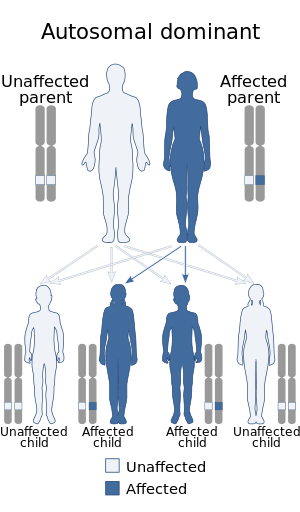
| Spondyloperipheral dysplasia has an autosomal dominant pattern of inheritance. | |
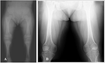
Spondyloperipheral dysplasia is an autosomal dominant[1] disorder of bone growth. The condition is characterized by flattened bones of the spine (platyspondyly) and unusually short fingers and toes (brachydactyly). Some affected individuals also have other skeletal abnormalities, short stature, nearsightedness (myopia), hearing loss, and mental retardation. Spondyloperipheral dysplasia is a subtype of collagenopathy, types II and XI.
Symptoms and signs
The clinical presentation of this condition is as follows:[2]
- Cleft palate
- Abnormality of vertebral endplates
- Abnormality of hip joint
- Myopia
- Cataract
Genetics
Spondyloperipheral dysplasia is one of a spectrum of skeletal disorders caused by mutations in the COL2A1 gene, located on chromosome 12q13.11-q13.2.[3] The protein made by this gene forms type II collagen, a molecule found mostly in cartilage and in the clear gel that fills the vitreous humour (the eyeball). Type II collagen is essential for the normal development of bones and other connective tissues (the tissues that form the body's supportive framework).
Mutations in the COL2A1 gene interfere with the assembly of type II collagen molecules. The protein made by the altered COL2A1 gene cannot be used to make type II collagen, resulting in a reduced amount of this type of collagen in the body. Instead of forming collagen molecules, the abnormal protein builds up in cartilage cells (chondrocytes). These changes disrupt the normal development of bones, leading to the signs and symptoms of spondyloperipheral dysplasia.
The disorder is believed to be inherited in an autosomal dominant manner.[1] This indicates that the defective gene responsible for the disorder is located on an autosome (chromosome 12 is an autosome), and only one copy of the defective gene is sufficient to cause the disorder, when inherited from a parent who has the disorder.
Diagnosis

The diagnosis for Spondyloperipheral dysplasia can be carried out via genetic testing[4]
Management
The management is per symptom that the individual is exhibiting[5]
References
- ↑ 1.0 1.1 Zabel, B.; Hilbert, K.; Stöß, H.; Superti-Furga, A.; Spranger, J.; Winterpacht, A. (May 1996). "A specific collagen type II gene (COL2A1) mutation presenting as spondyloperipheral dysplasia". American Journal of Medical Genetics. 63 (1): 123–128. doi:10.1002/(SICI)1096-8628(19960503)63:1<123::AID-AJMG22>3.0.CO;2-P. PMID 8723097.
- ↑ "Spondyloperipheral dysplasia | Genetic and Rare Diseases Information Center (GARD) – an NCATS Program". rarediseases.info.nih.gov. Archived from the original on 18 March 2021. Retrieved 2 August 2021.
- ↑ Online Mendelian Inheritance in Man (OMIM): 120140
- ↑ "Spondyloperipheral dysplasia-short ulna syndrome - Conditions - GTR - NCBI". www.ncbi.nlm.nih.gov. Archived from the original on 6 November 2020. Retrieved 2 August 2021.
- ↑ Spranger, Jürgen W.; Brill, Paula W.; Hall, Christine; Nishimura, Gen; Superti-Furga, Andrea; Unger, Sheila (25 October 2018). Bone Dysplasias: An Atlas of Genetic Disorders of Skeletal Development. Oxford University Press. p. 92. ISBN 978-0-19-062666-2. Archived from the original on 29 August 2021. Retrieved 2 August 2021.
This article incorporates public domain text from Spondyloperipheral dysplasia at NLM Genetics Home Reference
External links
- Spondyloperipheral dysplasia short ulna at NIH's Office of Rare Diseases
| Classification | |
|---|---|
| External resources |