Skin examination
| Skin examination | |
|---|---|
| Specialty | Dermatology |
Skin examination is an assessment of skin, hair, nails, and mouth, with the purpose of diagnosing a skin condition.[1] It is generally performed after taking a history.[2]
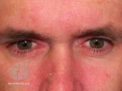
Distribution
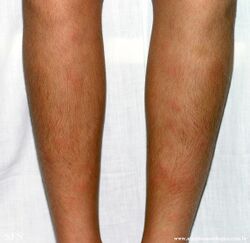
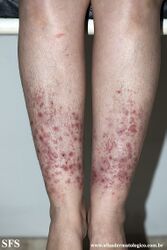
Symmetrical
A symmetrical distribution typically results from an internal cause, though not always.[1]
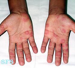
-
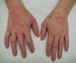
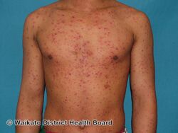
Symmetrical distribution in eczema
-
-

.jpg)
.jpg)
Unilateral
-
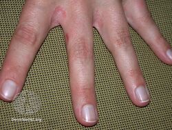
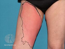
One hand affected in externally caused contact dermatitis
-
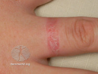
One finger affected due to contact allergy to gold
-
_(DermNet_NZ_dermatitis-icd-2510).jpg)
.jpg)
.jpg)
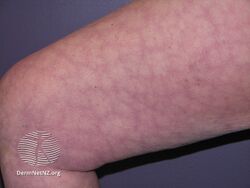
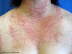
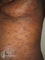
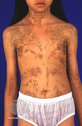
Widespread
-
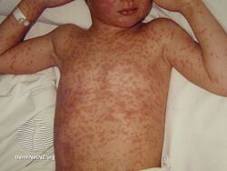
Widespread distribution in chicken pox with sparing of palms of hands
-
.jpg)
_(DermNet_NZ_viral-measles12).jpg)
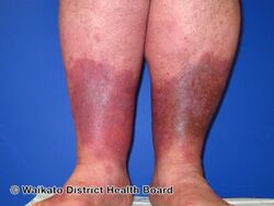
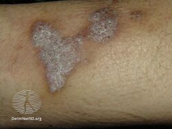
Site specific
-
Erythema nodosum (shins)
-
Pretibial Dominant Dystrophic Epidermolysis Bullosa (shins)
.jpg)
.jpg)
Pattern
Patterns include lace-like, and snake-like.[1]
-
Cutis marmorata (reticulate pattern)
-

Psoriasis (extensor surfaces)
-
-
Seborrhoeic dermatitis (alar groove and nasolabial fold)
-
Polymorphic light eruption in distribution of sun-exposed part of body
-
Photocontact dermatitis (sparing of covered part of body)
-
Pityriasis rosea (follows Langer's lines)
-
Epidermal nevus (follows lines of Blaschko)
.jpg)

.jpg)
.jpg)
.jpg)
.jpg)
.jpg)
.jpg)
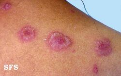
Shape
Skin lesion shapes include lines, spots, and bumps.[1]
-
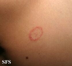
Tinea corporis (like a ring)
-

Psoriasis (like a coin)
-
Erythema multiforme (like a target)
-
Lichen plants (purplish, polygonal, Wickham striae)
-
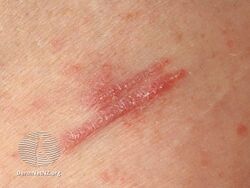
Koebner phenomenon in lichen planus (line)
.jpg)

.jpg)
.jpg)
.jpg)
See also
References
- ↑ 1.0 1.1 1.2 1.3 Tidman, Michael J. (2018). "14. The skin, hair and nails". Macleod's Clinical Examination (14th ed.). Edinburgh: Elsevier. pp. 283–292. ISBN 978-0-7020-6991-8. Archived from the original on 2024-06-01. Retrieved 2024-05-31.
- ↑ Buckley, David (2021). "2. History taking and examination". In Buckley, David; Pasquali, Paola (eds.). Textbook of Primary Care Dermatology. Switzerland: Springer. pp. 7–12. ISBN 978-3-030-29100-6. Archived from the original on 2024-06-10. Retrieved 2024-06-07.