User talk:QuackGuru/Sand A

From WikiProjectMed
Jump to navigation Jump to search

{{Db-u1|No longer needed.}} https://commons.wikimedia.org/wiki/Special:Watchlist

Requests

Discussion on requests

User:痛, I need your professional expertise for the specific requests below. Are you interested in working on any of these requests? Please let me know. Thanks.

Gif request

Create a gif from 15.5 to 18 seconds.

I request a gif from 15.5 to 18 seconds.

Montage for a new gif

This montage will be part of a new gif as part of a series of lung diagrams and other images. I request a montage like this with only four images. I suggest using the image with the text "Top half of montage image" for the top part of the montage. Then select two other images for the left side (Middle Left and Bottom Left) and one image for the right side.

I picked only four of the images above for the montage, starting with the "Top half of montage image".

Montage and gif 1

Completed montage

This montage is completed and will be used as the first montage for the new gif.

Montage and gif 2

Top half
Middle left
Bottom left
Right half (may need to be cropped to fit)
Make like this montage

Male and female name suggestion: File:Vape-cloud-montage-scaled 2.jpg

This montage will be used as the second montage for the gif. The first step is to create a new montage with the female and male images above.

Montage and gif 3

Top half
Middle left
Bottom left
Right half (may need to be cropped to fit)
Make like this montage

Females name suggestion: File:Vape-cloud-montage-scaled 3.jpg

This montage will be used as the third and last montage for the gif. The second step is to create a second montage with the female images above. Then, the final step is to create at least a five second gif for each montage while using all three montages.

Vaping lung disease outbreak map

Upload a new file under the name "File:2019–2020 vaping lung disease outbreak - fatalities.svg"

Various states need to be updated in the new map under a new file name.

  At least one death linked to a vaping product.
  At least two deaths linked to a vaping product.
  At least three deaths linked to a vaping product.
  At least four deaths linked to a vaping product.
  At least five deaths linked to a vaping product.
  At least six deaths linked to a vaping product.
  Hospitalizations but no confirmed deaths.

See: "Alabama, California (4)" For example, Alabama (AL) needs to be changed to orange. CA is already orange. No change needed for California (CA).

  At least four deaths linked to a vaping product.

See: "Connecticut (CT), Delaware (DE), District of Columbia (DC), Florida (FL) (2)"

  At least two deaths linked to a vaping product.

See: "Georgia (GA) (6)"

  At least six deaths linked to a vaping product.

See "Illinois (IL) (5)"

  At least five deaths linked to a vaping product.

See "Indiana (IN) (6)"

  At least six deaths linked to a vaping product.

See "Kansas (KS) (2)"

  At least two deaths linked to a vaping product.

See "Kentucky (KY), Louisiana (LA) (2)"

  At least two deaths linked to a vaping product.

See "Massachusetts (MA) (5)"

  At least five deaths linked to a vaping product.

See "Michigan (MI) (3)"

  At least three deaths linked to a vaping product.

See "Minnesota (MN) (3)"

  At least three deaths linked to a vaping product.

See "Mississippi (MS), Missouri (MO) (2)"

  At least two deaths linked to a vaping product.

See "Montana (MT), Nebraska (NE), New Jersey (NJ), New York (NY) (4)"

  At least four deaths linked to a vaping product.

See "Oregon (OR) (2)"

  At least two deaths linked to a vaping product.

See "Pennsylvania (PA), Rhode Island (RI), South Carolina (SC), Tennessee (TN) (2)"

  At least two deaths linked to a vaping product.

See "Texas (TX) (4)"

  At least four deaths linked to a vaping product.

See "Utah (UT), Virginia (VA) and Washington (WA) (2)"
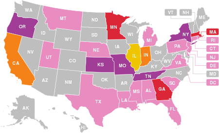
  At least two deaths linked to a vaping product.

Here is a link to a large blowup of this map. The link will make it easier to find each state in order to update each state and each color. Here is a link to a different map with each state spelled out.

69 deaths associated with the use of vaping products have been confirmed in the US, as of February 18, 2020[1] - this map shows states with confirmed fatalities.

  Indicates at least one death linked to a vaping product
  Indicates at least two deaths linked to a vaping product
  Indicates at least three deaths linked to a vaping product
  Indicates at least four deaths linked to a vaping product
  Indicates at least five deaths linked to a vaping product
  Indicates at least six deaths linked to a vaping product.
  Hospitalizations but no confirmed deaths.

For verification see, "Sixty-eight deaths have been confirmed in 29 states and the District of Columbia (as of February 18, 2020): Alabama, California (4), Connecticut, Delaware, District of Columbia, Florida (2), Georgia (6), Illinois (5), Indiana (6), Kansas (2), Kentucky, Louisiana (2), Massachusetts (5), Michigan (3), Minnesota (3), Mississippi, Missouri (2), Montana, Nebraska, New Jersey, New York (4), Oregon (2), Pennsylvania, Rhode Island, South Carolina, Tennessee (2), Texas (4), Utah, Virginia and Washington (2)."[2]

Requests

Discussion on requests

User:VulpesVulpes42, a picture can speak without making a word.

Discussion on requests

User:PawełMM, I need professional assistance with uploading and/or making modifications for several of the requests below.

Fundamentals of chemosensation

https://en.wikipedia.org/wiki/Taste#Basic_tastes

https://www.ncbi.nlm.nih.gov/pmc/articles/PMC9365686/#S2title Copy entire section.

Figure shows summary of three distinct sensory pathways involved in flavor perception. Taste, smell, and chemesthesis are integrated in the orbitofrontal cortex (OFC) to generate the unitary percept that we call flavor. Taste signals are carried by Cranial Nerves VII, IX, and X to the nucleus of the solitary tract (NST), which connects to the Ventral Posteromedial Nucleus (VPM) within the Thalamus. The VPM projects to the taste cortex located in the Insula. CN VII (the facial nerve) has two branches involved in taste: the chorda tympani (CT) innervates the anterior tongue while the greater superficial petrosal nerve (GSP) innervates the palate. Smell signals are carried by Cranial Nerve I through the cribriform plate to the olfactory bulb (OB), the Olfactory Cortex (OC), and then the Medial Dorsal Nucleus (MDN) within the Thalamus. Chemesthetic signals are carried by multiple cranial nerves, but for simplicity, only the trigeminal nerve (CN V) is shown here. Separate branches of the trigeminal nerve come together in the trigeminal ganglion (not shown), before descending to the spinal trigeminal nucleus (SpV) in the brain stem. From the SpV, signals ascend contralaterally to the VPM in the Thalamus, and then to the Somatosensory Cortex (SC). (Some anatomical landmarks in the brain have been omitted, and positions shown here may not be exact).
Figure shows summary of three distinct sensory pathways involved in flavor perception.[1] Taste, smell, and chemesthesis are integrated in the orbitofrontal cortex (OFC) to generate the unitary percept that we call flavor.[1] Taste signals are carried by Cranial Nerves VII, IX, and X to the nucleus of the solitary tract (NST), which connects to the Ventral Posteromedial Nucleus (VPM) within the Thalamus.[1] The VPM projects to the taste cortex located in the Insula.[1] CN VII (the facial nerve) has two branches involved in taste: the chorda tympani (CT) innervates the anterior tongue while the greater superficial petrosal nerve (GSP) innervates the palate.[1] Smell signals are carried by Cranial Nerve I through the cribriform plate to the olfactory bulb (OB), the Olfactory Cortex (OC), and then the Medial Dorsal Nucleus (MDN) within the Thalamus.[1] Chemesthetic signals are carried by multiple cranial nerves, but for simplicity, only the trigeminal nerve (CN V) is shown here.[1] Separate branches of the trigeminal nerve come together in the trigeminal ganglion (not shown), before descending to the spinal trigeminal nucleus (SpV) in the brain stem.[1] From the SpV, signals ascend contralaterally to the VPM in the Thalamus, and then to the Somatosensory Cortex (SC).[1] (Some anatomical landmarks in the brain have been omitted, and positions shown here may not be exact).[1]

By the early twentieth century, numerous researchers had recognized combined inputs from the taste, smell, and touch systems give rise to integrated percepts when we eat or drink.[1] In 1982, Rozin remarked that the word "flavor" best captures the combination of oral and olfactory sensations we perceive with ingestion of most foods, at least in English.[1] Today, most neuroscientists, sensory psychologists, and sensory and consumer scientists define flavor as the unitary percept which coalesces from the integration of smell, taste, and chemesthesis in the orbitofrontal cortex.[1] Despite this broad consensus, there remains some degree of confusion around these terms, regarding their colloquial and technical usage, even within medical professionals, so each of the three sensory modalities that contribute to flavor will be briefly detailed here.[1]

Olfaction (smell) occurs when we sense volatile chemical messages from the environment (via the nares) or from the oral cavity (through the back of the throat).[1] Odor active volatiles (i.e., odorants) activate specialized G-protein Coupled Receptors expressed in olfactory sensory neurons (OSNs) found near the top of the nasal cavity.[1] When an odorant binds to specialized receptor proteins expressed on the surface of OSNs, it initiates a transduction cascade which converts the chemical signal into an electrical signal.[1] The ensuing action potential is carried by the axon of the olfactory neuron through the cribriform plate, where the axons synapse onto second-order neurons in the olfactory bulb.[1] Because cell bodies of the OSNs sit at the top of the nasal cavity, below the cribriform plate, they are easily damaged by pollutants, viruses and toxins (including tobacco smoke).[1] However, OSNs are continually replaced, roughly every 30 days, which preserves function despite such environmental insults.[1] In contrast to other senses, smell is a dual sensory modality: that is, it occurs either orthonasally or retronasally and this affects where we localize the percept.[1] Ecologically speaking, orthonasal olfaction is an external sense focused on objects and information in the environment, while retronasal olfaction is an internally focused sense where volatiles that reach the olfactory epithelium via the pharyx during chewing or swallowing are perceived as being present in the mouth.[1]

Gustation (taste) occurs when non-volatile chemical stimuli dissolve in saliva and contact specialized taste receptor cells (TRCs) found in the tongue, soft palate and throat.[1] Unlike the OSNs mentioned above, the TRCs are not neurons—rather, they are specialized epithelial cells which must communicate with neurons to project a signal centrally.[1] Taste aids organisms in perception of nutrients and toxins, driving ingestion via affective responses.[1] The widely accepted prototypical taste qualities are sweet, salty, sour, bitter, and savory/umami (the meaty taste of certain amino acids).[1] Non-sweet starch taste, fatty acid taste (oleogustus), metallic taste, and astringent may also be distinct taste qualities, but the case for each is less clear and their inclusion as distinct qualities is still actively debated.[1] Individuals vary widely in terms of taste perception, due in part to genetic variation.[1] Such differences are potentially important for nicotine research, and are discussed more below.[1]

Chemesthesis is the sensibility that results from chemical stimulation of somatosensory nerves; that is, it can be thought of as chemically initiated touch.[1] Chemesthetic stimuli have a range of perceptual qualities, including the tingling elicited by carbonation, the burn from chili peppers, the burn from horseradish, the mechanical buzzing from Sichuan Buttons, and best known to tobacco researchers, the cooling from menthol.[1] As chemesthetic stimuli are known to trigger cough reflexes, they have strong relevance to e-cigarettes, especially given the importance of irritation or throat hit to e-cigarette liking and appeal.[1] Extensive discussions of menthol as it relates to use of nicotine containing products are covered in detail elsewhere, so comments below will be restricted to specific aspects related to narrowly to chemosensation.[1]

Notably, the classical assumption that nicotine is itself bitter is almost certainly in error.[1] Rather, three distinct and complementary lines of evidence suggest nicotine gives rise to chemesthetic sensations, rather than bitterness per se.[1] First, in heterologous expression systems, nicotine does not activate any known bitter taste receptor, but it does activate TRPA1, a receptor activated by ligands like cinnamaldehyde or allyl isothiocyanate (AITC) that impart the pungency of cinnamon and wasabi, respectively.[1] Second, electrophysiology data from rats and psychophysical data from humans each indicate nicotine is a chemesthetic stimulus.[1] Third, close reading of very old literature suggests a widely cited 1959 source for the widespread claim that nicotine is bitter in turn leads back to an earlier paper from 1885.[1] Critically, if one reads the original source from 1885, the authors explicitly write "nicotine does not trigger a taste sensation," noting that if the concentration is increased, it produces "a stinging sensation, which is not, strictly speaking, a taste sensation, but tactile".[1] This caveat notwithstanding, combustible tobacco smoke certainly gives rise to bitter sensations from one of the hundreds of other compounds found in smoke, but strictly speaking it does not seem such bitterness can be directly attributable to nicotine.[1] Regarding e-cigarettes, participants report bitterness in multiple studies, but the source of this bitterness remains unknown.[1]

[1]

References

  1. 1.00 1.01 1.02 1.03 1.04 1.05 1.06 1.07 1.08 1.09 1.10 1.11 1.12 1.13 1.14 1.15 1.16 1.17 1.18 1.19 1.20 1.21 1.22 1.23 1.24 1.25 1.26 1.27 1.28 1.29 1.30 1.31 1.32 1.33 1.34 1.35 1.36 1.37 1.38 1.39 1.40 1.41 Hayes, John E.; Baker, Allison N. (27 July 2022). "Flavor science in the context of research on electronic cigarettes". Frontiers in Neuroscience. 16. doi:10.3389/fnins.2022.918082. PMC 9365686. PMID 35968379. This article incorporates text by John E Hayes and Allison N Baker available under the CC BY 4.0 license.

Blood-brain barrier

https://en.wikipedia.org/wiki/Cocaine

Schematic representation showing drug-induced loss of blood brain barrier (BBB) permeability and the associated neurodegeneration. The neurovascular unit and the BBB are affected by various drugs of abuse, which alter vessel permeability via disruption of tight junction proteins complexes (junction adhesion molecules, endothelial cell-selective adhesion molecules, occludins, and claudins), transport systems, and intracellular signaling. BBB disruption, which affects immune cell transmigration and neuroinflammation and contributes to an imbalanced redox system, affects the brain’s microenvironment and homeostasis, leading to neurotoxicity.
Schematic representation showing drug-induced loss of blood-brain barrier (BBB) permeability and the associated neurodegeneration.[1] The neurovascular unit and the BBB are affected by various drugs of abuse, which alter vessel permeability via disruption of tight junction proteins complexes (junction adhesion molecules, endothelial cell-selective adhesion molecules, occludins, and claudins), transport systems, and intracellular signaling.[1] BBB disruption, which affects immune cell transmigration and neuroinflammation and contributes to an imbalanced redox system, affects the brain’s microenvironment and homeostasis, leading to neurotoxicity.[1]

The World Drug Report estimates that worldwide, 18.8 million people used cocaine in 2014.[1] In 2016, the National Institute on Drug Abuse reported an age-adjusted cocaine-mediated death rate of 52.4% in the US.[1] Cocaine is a highly addictive stimulant that restricts dopamine and monoamine reuptake through dopamine transporter (DAT) antagonism.[1] Monoamine oxidase inhibition leads to imbalanced free-radical production, which generates oxidative stress and neuroinflammation.[1] Continuous cocaine administration has been shown to contribute to a 50% increase in blood-brain barrier (BBB) permeability, with a concomitant decrease in trans endothelial electrical resistance (TEER) due to basement membrane and neurovascular capillary disruption, due to up-regulated matrix metalloproteinase (MMP) and tumor necrosis factor (TNF-α) expression.[1] Moreover, TJ protein loss and alteration, specifically decreased JAM-2 and zonula occludens-1 (ZO-1) levels, are characteristic of cocaine transit across the BBB.[1] CCL2 (C-C motif chemokine ligand-2) and CCR2 (C-C motif chemokine receptor-2) expression upregulation has also been reported.[1] Cocaine use affects intercellular junctions and causes cell ruffling, which contributes to increased permeability and decreased TEER values across BBB monolayers.[1]

An alternate pathway for cocaine-induced BBB permeability alteration involves platelet-derived growth factor (PDGF) intermediates.[1] Cocaine binding to sigma receptors evokes a proteolytic signal cascade that initiates PDGF-B chain assembly, a fundamental intermediate for increased membrane permeability that inhibits store-operated calcium entry.[1] Moreover, cocaine binding to sigma receptors has been associated with dopamine uptake inhibition and enhanced dopamine release that neutralizes the effects of antibody reversal on increased PDGF expression.[1] In rats, chronic cocaine exposure has been shown to increase BBB permeability in the hippocampus and striatum, suggesting that the hippocampus could be affected by glial and cytokine migration without significant changes in cortical or cerebellar permeability.[1] Furthermore, it has been recently revealed that acute cocaine administration alters BBB permeability and may increase neurotoxicity in free-moving rats.[1]

Astrocytes have complex morphologies involving extensive processes that communicate within the neurovascular unit and maintain the BBB.[1] Cocaine exposure potentiates aberrant astroglial responses in cellular and animal models, which leads to loss of BBB integrity and function.[1] Other studies have reported cocaine-induced neuroinflammation and BBB disruption mediated by the activation of brain microglial cells to secrete several cytokines, chemokines, and other neurotoxic factors.[1] Cocaine upregulates these inflammatory mediators and cell adhesion molecules, including intercellular adhesion molecule-1, vascular cell adhesion molecule, and activated leukocyte cell adhesion molecule in the BBB endothelium.[1]

Previous in vitro findings have shown that exposure of pericytes to cocaine upregulates pro-inflammatory cytokines [TNF-α, interleukin (IL)-1β, and IL-6] in both intracellular and extracellular compartments.[1] In addition, cocaine activates the Src–PDGFR-β–NF-κB pathway, which enhances CXCL10 [chemokine (C-X-C motif) ligand-1] secretion.[1] This causes increased neuroinflammation in human brain vascular pericytes, which further leads to neurovascular unit disruption and immune cell transmigration across the BBB.[1]

Blood-brain barrier

https://en.wikipedia.org/wiki/Methamphetamine

Schematic representation showing drug-induced loss of blood brain barrier (BBB) permeability and the associated neurodegeneration. The neurovascular unit and the BBB are affected by various drugs of abuse, which alter vessel permeability via disruption of tight junction proteins complexes (junction adhesion molecules, endothelial cell-selective adhesion molecules, occludins, and claudins), transport systems, and intracellular signaling. BBB disruption, which affects immune cell transmigration and neuroinflammation and contributes to an imbalanced redox system, affects the brain’s microenvironment and homeostasis, leading to neurotoxicity.
Schematic representation showing drug-induced loss of blood-brain barrier (BBB) permeability and the associated neurodegeneration.[1] The neurovascular unit and the BBB are affected by various drugs of abuse, which alter vessel permeability via disruption of tight junction proteins complexes (junction adhesion molecules, endothelial cell-selective adhesion molecules, occludins, and claudins), transport systems, and intracellular signaling.[1] BBB disruption, which affects immune cell transmigration and neuroinflammation and contributes to an imbalanced redox system, affects the brain’s microenvironment and homeostasis, leading to neurotoxicity.[1]

Methamphetamine is a highly addictive and illicit psychostimulant and is the second most widely abused drug in the US.[1] It adversely affects brain homeostasis through blood-brain barrier (BBB) dysfunction and hyperthermia.[1] Its high lipophilicity allows for rapid and comprehensive transmigration across the BBB.[1] Methamphetamine binding to the DAT induces reversal transport of norepinephrine, serotonin (5HT), and dopamine, which causes their excessive release into the synapse.[1] Moreover, it inhibits monoamine reuptake that leads to post-synaptic cleft stimulation.[1] Chronic methamphetamine administration causes irreversible impairment of serotonin and dopamine transport into synaptic terminals in various brain regions, especially in the hippocampus.[1]

Various methamphetamine dosing paradigms significantly disturb endothelial TJ assembly by inducing downregulation, fragmentation, or redistribution of major TJ proteins, including claudin-5 and ZO-1, which are mediated by MMP-1 and MMP-9 peptidases.[1] This leads to reduced endothelial barrier tightness and increased BBB paracellular permeability.[1] Moreover, repeated intravenous methamphetamine administration downregulates TJ proteins, which causes glutathione depletion and increases endothelial reactive oxygen species (ROS) levels.[1] This triggers actin polymerization that possibly involves activation of actin-related protein 2/3 complex or myosin light chain kinase and its downstream target RhoA.[1] In mice, research has shown that methamphetamine-induced glucose transporter and uptake downregulation is an important causative factor for BBB integrity loss.[1] Further, methamphetamine reduces TJ protein expression, rearranges the F-actin cytoskeleton, and increases BBB permeability through Rho-associated protein kinase-dependent pathway activation in the frontal lobes and isolated primary microvascular endothelial cells.[1]

Other neurotoxicity mechanisms have also been suggested, including the methamphetamine-induced increase in reactive oxidative stress and ROS levels, which activate myosin light chain protein kinase, thereby reducing TJ protein expression.[1] Additionally, methamphetamine-induced TJ protein downregulation and resulting BBB integrity disruption may involve activation of NF-κB transcription and pro-inflammatory cytokines (TNF-α) in BBB endothelial cells.[1] Methamphetamine transit across the BBB damages the nucleus accumbens shell region and prefrontal cortex and causes hyperthermia, neuroinflammation, and brain edema.[1] Studies have reported methamphetamine-induced pericyte migration via sigma-1 receptor activation, p53 upregulated modulator of apoptosis expression, and downstream mitogen-activated protein kinase and Akt/PI3K pathways in C3H/10T1/2 cells, leading to BBB dysfunction.[1] Methamphetamine-activated microglia and astrocytes in the neurovascular unit may promote neurotoxicity and astroglial reactivity and induces BBB integrity loss.[1] In addition, methamphetamine increases the expression of the glial fibrillary acidic protein, σ1 receptors, TNF-α, IL-6, and IL-8 in mouse and rat astrocytes.[1] This leads to methamphetamine-induced inflammation in microglial cells where increased TNF-α release can activate BBB endothelium, which increases transmigration of circulating leukocytes through the leaky BBB.[1]

Blood-brain barrier

https://en.wikipedia.org/wiki/Morphine

Schematic representation showing drug-induced loss of blood brain barrier (BBB) permeability and the associated neurodegeneration. The neurovascular unit and the BBB are affected by various drugs of abuse, which alter vessel permeability via disruption of tight junction proteins complexes (junction adhesion molecules, endothelial cell-selective adhesion molecules, occludins, and claudins), transport systems, and intracellular signaling. BBB disruption, which affects immune cell transmigration and neuroinflammation and contributes to an imbalanced redox system, affects the brain’s microenvironment and homeostasis, leading to neurotoxicity.
Schematic representation showing drug-induced loss of blood-brain barrier (BBB) permeability and the associated neurodegeneration.[1] The neurovascular unit and the BBB are affected by various drugs of abuse, which alter vessel permeability via disruption of tight junction proteins complexes (junction adhesion molecules, endothelial cell-selective adhesion molecules, occludins, and claudins), transport systems, and intracellular signaling.[1] BBB disruption, which affects immune cell transmigration and neuroinflammation and contributes to an imbalanced redox system, affects the brain’s microenvironment and homeostasis, leading to neurotoxicity.[1]

Opioids are widely used analgesics that bind with opioid and/or toll-like receptors (TLR) in the CNS.[1] Transcellular solute and xenobiotic transport across the blood-brain barrier (BBB) is selectively controlled by the local influx and efflux transporters, including ATP-binding cassette (ABC), P-glycoprotein (P-gp, ABCB1), breast cancer resistance protein (ABCG2), multidrug resistance-associated proteins (ABCC) transporters, and solute carrier transporters.[1] Among the four central opioid receptor families [mu (μ), delta (δ), kappa (κ), and opioid receptor like-1 (ORL1) receptor], μ-opioid receptors are primarily responsible for the analgesic effects.[1] Microvascular endothelial cells have high affinity and specific opiate binding sites that mediate morphine’s effects on the CNS.[1]

Morphine exerts its effects by directly acting on the CNS with its illicit use leading to tolerance and drug dependence.[1] Drug transmigration is essential to psychological dependence.[1] Morphine alters BBB homeostasis and permeability through pro-inflammatory cytokine activity, intracellular calcium release dysregulation, and myosin light chain protein kinase activation, which results in ROS-mediated neurotoxicity.[1]

P-gp limits the net transport of several foreign substrates into the brain through active unidirectional efflux.[1] This transporter regulates foreign-agent pharmacokinetics in the brain by inhibiting or augmenting their movement across the BBB, which restrains morphine entry into the brain.[1] Moreover, P-gp attenuates morphine-induced migratory properties and transcriptional effects.[1] Acute morphine treatment inhibits P-gp expression, which increases morphine uptake in the brain, which modifies the acute analgesic and locomotive morphine effects and selectively alters critical transcriptional responses in the nucleus accumbens.[1] This indicates that the transporter system significantly contributes to mediating BBB integrity and permeability of carrier mediated transport.[1]

Blood-brain barrier

https://en.wikipedia.org/wiki/Heroin

Schematic representation showing drug-induced loss of blood brain barrier (BBB) permeability and the associated neurodegeneration. The neurovascular unit and the BBB are affected by various drugs of abuse, which alter vessel permeability via disruption of tight junction proteins complexes (junction adhesion molecules, endothelial cell-selective adhesion molecules, occludins, and claudins), transport systems, and intracellular signaling. BBB disruption, which affects immune cell transmigration and neuroinflammation and contributes to an imbalanced redox system, affects the brain’s microenvironment and homeostasis, leading to neurotoxicity.
Schematic representation showing drug-induced loss of blood-brain barrier (BBB) permeability and the associated neurodegeneration.[1] The neurovascular unit and the BBB are affected by various drugs of abuse, which alter vessel permeability via disruption of tight junction proteins complexes (junction adhesion molecules, endothelial cell-selective adhesion molecules, occludins, and claudins), transport systems, and intracellular signaling.[1] BBB disruption, which affects immune cell transmigration and neuroinflammation and contributes to an imbalanced redox system, affects the brain’s microenvironment and homeostasis, leading to neurotoxicity.[1]

There has been a rapid increase in opioid abuse in the US with approximately 580 new heroin users every day.[1] Deaths resulting from opiate overdose, including pain relievers and heroin, increased by 200% between 2000 and 2014.[1] Heroin can be reversibly metabolized into morphine; upon selective transmigration across the blood-brain barrier (BBB), heroin is transformed into morphine and metabolized into 6-monoacetylmorphine (6-MAM).[1] The superior heroin lipophilicity allows faster transit across the BBB than morphine.[1] The acetylation of both hydroxyl groups while synthesizing heroin increases its BBB penetration rate by 100-fold, which could contribute to its high addictive potential.[1] These addictive properties are regulated by the μ-opioid receptor (MOR), which mediates the rewarding effects of heroin.[1] A recent study reported that 6-MAM has a higher affinity for μ-opioid receptor G-protein activation than morphine.[1]

Heroin's effects indirectly involve its metabolites (morphine and 6-MAM) that act as substrates in P-gp membrane regulation.[1] Upon heroin transition into the brain, it has a higher synthesized concentration than morphine.[1] This suggests that the metabolite is the primary effector of the detrimental effects of heroin on the BBB.[1] In the extracellular brain fluid, these metabolites bind and activate MORs, which regulates crucial neurological automatic processes.[1] P-gp inhibition at the BBB acutely disrupts the BBB permeability and selectivity in the nucleus accumbens.[1] Moreover, increased levels of these metabolites in the brain downregulate TJ protein expression, especially ZO-1, which increases BBB permeability.[1] Contrastingly, there have been reports of increased JAM-2 TJ protein expression.[1]

Blood-brain barrier

https://en.wikipedia.org/wiki/Alcohol_and_health

Schematic representation showing drug-induced loss of blood brain barrier (BBB) permeability and the associated neurodegeneration. The neurovascular unit and the BBB are affected by various drugs of abuse, which alter vessel permeability via disruption of tight junction proteins complexes (junction adhesion molecules, endothelial cell-selective adhesion molecules, occludins, and claudins), transport systems, and intracellular signaling. BBB disruption, which affects immune cell transmigration and neuroinflammation and contributes to an imbalanced redox system, affects the brain’s microenvironment and homeostasis, leading to neurotoxicity.
Schematic representation showing drug-induced loss of blood-brain barrier (BBB) permeability and the associated neurodegeneration.[1] The neurovascular unit and the BBB are affected by various drugs of abuse, which alter vessel permeability via disruption of tight junction proteins complexes (junction adhesion molecules, endothelial cell-selective adhesion molecules, occludins, and claudins), transport systems, and intracellular signaling.[1] BBB disruption, which affects immune cell transmigration and neuroinflammation and contributes to an imbalanced redox system, affects the brain’s microenvironment and homeostasis, leading to neurotoxicity.[1]

Alcohol is a widely used recreational drug responsible for 5.3% of deaths worldwide.[1] In the US, there are 23 million alcohol addicts with 88,000 people dying from alcohol use disorder.[1] Alcohol acts on neurotransmitter receptors, including GABA, glutamate, and dopamine, with each receptor contributing to various physiologic effects, with chronic alcohol administration increasing tolerance and addiction.[1] Further, occasional alcohol consumption could lead to alcohol use disorder due to addiction and tolerance.[1] Regular and excessive alcohol consumption causes brain injury, white matter loss, reduced brain volume, and neuronal loss associated with the BBB.[1] Moreover, gray matter loss is positively correlated with years of alcohol abuse.[1] Chronic alcohol abuse induces neuroplastic changes and loss of neural circuit structure and strength.[1]

The brains of individuals with alcohol dependence have increased proinflammatory cytokines, chemokines, microglial markers, and inflammasome proteins.[1] Inflammatory cytokine and ROS activation contributes to BBB integrity disruption in TLR4-knockout mice.[1] Further, postmortem alcoholic brains have shown increased TLR2, TLR3, and TLR4 expression in the orbitofrontal cortex, which correlates with BBB integrity loss.[1] Moreover, they indicate that chronic alcohol intake increases TJ and neuroinflammatory protein (ERK1/2 and p-38) degradation, which may promote leukocyte brain infiltration.[1]

Brain microvascular endothelial cells (BMVEC) are interconnected with TJ to form a tight monolayer in the BBB.[1] Exposure of BMVEC to alcohol increases oxidative stress through myosin light chain and TJ protein phosphorylation.[1] This leads to decreased TEER and increased leukocyte migration across the BBB.[1] Further, alcohol induces BBB dysfunction and neuroinflammation through MMP-3/9 activation and angiogenesis (VEGF)-A/VEGFR-2) impairment in primary endothelial cells in the brain.[1] Ethanol (EtOH) disrupts BBB integrity via endothelial transient receptor potential (TRP) channels, which affects the intracellular Ca2+ and Mg2+ dynamics.[1] This increases endothelial permeability and alters inflammatory responses.[1] EtOH-mediated TRPM7 expression downregulation causes BBB dysfunction and endothelium integrity loss.[1] Overall, TRP channels are involved in alcohol-mediated BBB dysfunction.[1]

Figure 1

=={{int:filedesc}}==

⧼wm-license-information-description⧽ Transclusion error: {{En}} is only for use in File namespace. Use {{lang-en}} or {{in lang|en}} instead.
⧼wm-license-information-date⧽
⧼wm-license-information-source⧽ https://www.frontiersin.org/journals/public-health/articles/10.3389/fpubh.2019.00287/full
⧼wm-license-information-author⧽ Nadja Mallock, Elke Pieper, Christoph Hutzler, Frank Henkler-Stephani, and Andreas Luch

{{cc-by-4.0}}

[[Category:Heated tobacco products]]

CC BY 4.0 DEED Attribution 4.0 International[3]

Copyright © 2019 Mallock, Pieper, Hutzler, Henkler-Stephani and Luch. This is an open-access article distributed under the terms of the Creative Commons Attribution License (CC BY). The use, distribution or reproduction in other forums is permitted, provided the original author(s) and the copyright owner(s) are credited and that the original publication in this journal is cited, in accordance with accepted academic practice. No use, distribution or reproduction is permitted which does not comply with these terms.[4]

Authors: Nadja Mallock, Elke Pieper, Christoph Hutzler, Frank Henkler-Stephani, and Andreas Luch

Received: 26 March 2019; Accepted: 20 September 2019; Published: 10 October 2019.

Figure 1

Temperature zones in a combustible cigarette (A) in comparison to different Heated Tobacco Products (B).

https://www.ncbi.nlm.nih.gov/pmc/articles/PMC6795920/

[2]

Suggested file name:

File:Temperature zones in a combustible cigarette in comparison to different heated tobacco products.jpg

The illustration shows the temperature zones in a combustible cigarette (A) in comparison to different heated tobacco products (B). Heated tobacco products differ widely in product design and temperatures applied to the tobacco. In some devices the tobacco is heated up to 350 °C via an electrical heating source or different sources like carbon, whereas in other devices aerosol is passed through the tobacco and extracts compounds including flavors and nicotine at lower temperatures. Three different device designs which are currently present on the market are displayed here. These products contain real tobacco.
The illustration shows the temperature zones in a combustible cigarette (A) in comparison to different heated tobacco products (B).[2] Heated tobacco products differ widely in product design and temperatures applied to the tobacco.[2] In some devices the tobacco is heated up to 350 °C via an electrical heating source or different sources like carbon, whereas in other devices aerosol is passed through the tobacco and extracts compounds including flavors and nicotine at lower temperatures.[2] Three different device designs which are currently present on the market are displayed in here.[2] These products contain real tobacco.[2]

References

  1. 1.00 1.01 1.02 1.03 1.04 1.05 1.06 1.07 1.08 1.09 1.10 1.11 1.12 1.13 1.14 1.15 1.16 1.17 1.18 1.19 1.20 1.21 1.22 1.23 1.24 1.25 1.26 1.27 1.28 1.29 1.30 1.31 1.32 1.33 1.34 1.35 1.36 1.37 1.38 1.39 1.40 1.41 1.42 1.43 1.44 1.45 1.46 1.47 1.48 1.49 1.50 1.51 1.52 1.53 1.54 1.55 1.56 1.57 1.58 1.59 1.60 1.61 1.62 1.63 1.64 1.65 1.66 1.67 1.68 1.69 1.70 1.71 1.72 1.73 1.74 1.75 1.76 1.77 1.78 1.79 1.80 1.81 1.82 1.83 1.84 1.85 1.86 1.87 1.88 1.89 1.90 1.91 1.92 1.93 1.94 1.95 1.96 1.97 1.98 Pimentel, Emely; Sivalingam, Kalaiselvi; Doke, Mayur; Samikkannu, Thangavel (21 May 2020). "Effects of Drugs of Abuse on the Blood-Brain Barrier: A Brief Overview". Frontiers in Neuroscience. 14. doi:10.3389/fnins.2020.00513. PMC 7326150. PMID 32670001. This article incorporates text by Emely Pimentel, Kalaiselvi Sivalingam, Mayur Doke, and Thangavel Samikkannu available under the CC BY 4.0 license.
  2. 2.0 2.1 2.2 2.3 2.4 2.5 Mallock, Nadja; Pieper, Elke; Hutzler, Christoph; Henkler-Stephani, Frank; Luch, Andreas (10 October 2019). "Heated Tobacco Products: A Review of Current Knowledge and Initial Assessments". Frontiers in Public Health. 7. doi:10.3389/fpubh.2019.00287. PMC 6795920. PMID 31649912. This article incorporates text by Nadja Mallock, Elke Pieper, Christoph Hutzler, Frank Henkler-Stephani, and Andreas Luch available under the CC BY 4.0 license.

FIGURE 2, FIGURE 3 and FIGURE 6

Licensing for the content is {{cc-by-4.0}}.

FIGURE 2

=={{int:filedesc}}==

⧼wm-license-information-description⧽ Transclusion error: {{En}} is only for use in File namespace. Use {{lang-en}} or {{in lang|en}} instead.
⧼wm-license-information-date⧽
⧼wm-license-information-source⧽ https://www.frontiersin.org/journals/pharmacology/articles/10.3389/fphar.2023.1200269/full
⧼wm-license-information-author⧽ Eric Fordjour, Charles F. Manful, Albert A. Sey, Rabia Javed, Thu Huong Pham, Raymond Thomas, and Mumtaz Cheema

{{cc-by-4.0}}

[[Category:Category:Cannabis]]


FIGURE 3

=={{int:filedesc}}==

⧼wm-license-information-description⧽ Transclusion error: {{En}} is only for use in File namespace. Use {{lang-en}} or {{in lang|en}} instead.
⧼wm-license-information-date⧽
⧼wm-license-information-source⧽ https://www.frontiersin.org/journals/pharmacology/articles/10.3389/fphar.2023.1200269/full
⧼wm-license-information-author⧽ Eric Fordjour, Charles F. Manful, Albert A. Sey, Rabia Javed, Thu Huong Pham, Raymond Thomas, and Mumtaz Cheema

{{cc-by-4.0}}

[[Category:Category:Cannabis]]

FIGURE 6

=={{int:filedesc}}==

⧼wm-license-information-description⧽ Transclusion error: {{En}} is only for use in File namespace. Use {{lang-en}} or {{in lang|en}} instead.
⧼wm-license-information-date⧽
⧼wm-license-information-source⧽ https://www.frontiersin.org/journals/pharmacology/articles/10.3389/fphar.2023.1200269/full
⧼wm-license-information-author⧽ Eric Fordjour, Charles F. Manful, Albert A. Sey, Rabia Javed, Thu Huong Pham, Raymond Thomas, and Mumtaz Cheema

{{cc-by-4.0}}

[[Category:Category:Cannabis]]

See https://www.ncbi.nlm.nih.gov/pmc/articles/PMC10308385/#s1title

See https://www.frontiersin.org/journals/pharmacology/articles/10.3389/fphar.2023.1200269/full

Authors: Eric Fordjour, Charles F. Manful, Albert A. Sey, Rabia Javed, Thu Huong Pham, Raymond Thomas, and Mumtaz Cheema

Received: 05 April 2023; Accepted: 30 May 2023; Published: 15 June 2023.

FIGURE 2

The name of Cannabis in some popular languages.

FIGURE 3

Parts of Cannabis plants and products.

FIGURE 6

Pharmacological potential of Cannabis.

Copyright © 2023 Fordjour, Manful, Sey, Javed, Pham, Thomas and Cheema. This is an open-access article distributed under the terms of the Creative Commons Attribution License (CC BY). The use, distribution or reproduction in other forums is permitted, provided the original author(s) and the copyright owner(s) are credited and that the original publication in this journal is cited, in accordance with accepted academic practice. No use, distribution or reproduction is permitted which does not comply with these terms.[5]

[1]

References

  1. Fordjour, Eric; Manful, Charles F.; Sey, Albert A.; Javed, Rabia; Pham, Thu Huong; Thomas, Raymond; Cheema, Mumtaz (15 June 2023). "Cannabis: a multifaceted plant with endless potentials". Frontiers in Pharmacology. 14. doi:10.3389/fphar.2023.1200269. PMC 10308385. PMID 37397476. {{cite journal}}: Check |pmc= value (help) This article incorporates text by Eric Fordjour, Charles F. Manful, Albert A. Sey, Rabia Javed, Thu Huong Pham, Raymond Thomas, and Mumtaz Cheema available under the CC BY 4.0 license.

Figure 1

Licensing for the content is {{cc-by-4.0}}.

=={{int:filedesc}}== {{Information |description={{en|1=Digital necrosis }} |date=2017-01-17 |source=https://www.panafrican-med-journal.com/content/article/26/53/full/ |author=Naoual El Omri, Rachid El Jaoudi, Fadwa Mekouar, Mohammed Jira, Youssef Sekkach, Taoufik Amezyane, and Driss Ghafir |permission= |other versions= }} =={{int:license-header}}== {{cc-by-4.0}}

[[Category:Category:Cannabis]]

All articles published in this journal are Open Access and distributed under the terms of the Creative Commons Attribution 4.0 International (CC BY 4.0).

See https://www.panafrican-med-journal.com/content/article/26/53/full/

Also see https://www.panafrican-med-journal.com/content/article/26/53/full/figure.php?FigId=1

Figure 1 Digital necrosis

[1]

References

  1. Naoual, El Omri; Rachid, El Jaoudi; Fadwa, Mekouar; Mohammed, Jira; Youssef, Sekkach; Taoufik, Amezyane; Driss, Ghafir (2017). "Cannabis arteritis". Pan African Medical Journal. 26. doi:10.11604/pamj.2017.26.53.11694. PMC 5398854. PMID 28451030. This article incorporates text by Naoual El Omri, Rachid El Jaoudi, Fadwa Mekouar, Mohammed Jira, Youssef Sekkach, Taoufik Amezyane, and Driss Ghafir available under the CC BY 4.0 license.

Figure 5.

Content is in the public domain.

Click on "Information & Authors"[6] at the right-hand side.

HISTORY

Received: 4 March 2022

Revision received: 29 June 2022

Accepted: 28 July 2022

Published online: 14 September 2022

See under Copyright and License information:

EHP is an open-access journal published with support from the National Institute of Environmental Health Sciences, National Institutes of Health. All content is public domain unless otherwise noted.[7]

Authors: Naoual El Omri, Rachid Eljaoudi, Fadwa Mekouar, Mohammed Jira, Youssef Sekkach, Taoufik Amezyane, and Driss Ghafir

Figure 5.

Choropleth map showing the number of reported incidents of contaminated cannabis outbreaks and recalls in each jurisdiction between 1 June 2020 and 31 October 2021. News reports are searched using Google News, Newsbank, and Access World News. Eight of these 14 recalls were caused by microbial contamination. All news reports are listed in Supplemental Material, “Identified Regulatory Documents, Public Health Reports, Cannabis Testing Reports, and News Reports.” Note: AK, Alaska; AL, Alabama; AR, AR, Arkansas; AZ, Arizona; CA, California; CO, Colorado; CT, Connecticut; DE, Delaware; FL, Florida; HI, Hawaii; IL, Illinois; LA, Louisiana; MA, Massachusetts; MD, Maryland; ME, Maine; MI, Michigan; MN, Minnesota; MO, Missouri; MT, Montana; ND, North Dakota; NH, New Hampshire; NJ, New Jersey; NM, New Mexico; NV, Nevada; NY, New York; OH, Ohio; OR, Oregon; PA, RI, Rhode Island; UT, Utah; VA, Virginia; VT, Vermont; WA, Washington; WV, West Virginia.

[1]

References

  1. Jameson, Laura E.; Conrow, Kendra D.; Pinkhasova, Dorina V.; Boulanger, Haleigh L.; Ha, Hyunji; Jourabchian, Negar; Johnson, Steven A.; Simeone, Michael P.; Afia, Iniobong A.; Cahill, Thomas M.; Orser, Cindy S.; Leung, Maxwell C.K. (September 2022). "Comparison of State-Level Regulations for Cannabis Contaminants and Implications for Public Health". Environmental Health Perspectives. 130 (9). doi:10.1289/EHP11206. PMC 9472674. PMID 36102653.Public Domain This article incorporates text from this source, which is in the public domain.

New psychoactive substances by effect group

Upload "New psychoactive substances by effect group"

Content is in the public domain.

Date: 23 October 2023

Author: Stacy Lu

See https://nida.nih.gov/news-events/nida-asks/can-science-keep-up-with-designer-drugs

The data was obtained from the United Nations Office on Drugs and Crime, Early Warning Advisory on NPS. However, the National Institute on Drug Abuse created a new pie chart and used different colors.

See here for the previous pie chart on the United Nations Office on Drugs and Crime's website.

[1]

References

  1. Lu, Stacy (23 October 2023). "Can science keep up with designer drugs?". National Institute on Drug Abuse.

Figure

Upload Figure which states, Marijuana-Related Emergency Department (ED) Visits among Adolescents Aged 15 to 17, by Gender: 2005 and 2010

Content is in the public domain. See "The Data Spotlight may be copied without permission. Citation of the source is appreciated. Find this report and those on similar topics online at http://www.samhsa.gov/data/."

[1]

Figure 3

Licensing for the content is {{cc-by-4.0}}.

=={{int:filedesc}}==

⧼wm-license-information-description⧽ Transclusion error: {{En}} is only for use in File namespace. Use {{lang-en}} or {{in lang|en}} instead.
⧼wm-license-information-date⧽
⧼wm-license-information-source⧽ https://www.mdpi.com/1424-8247/14/10/965
⧼wm-license-information-author⧽ Vidyasagar Naik Bukke, Moola Archana, Rosanna Villani, Gaetano Serviddio, and Tommaso Cassano

=={{int:license-header}}==

{{cc-by-4.0}}

[[Category:Category:Cannabis]]

Figure 3

Toxicity of synthetic cannabinoids and their adverse effects.

https://www.mdpi.com/1424-8247/14/10/965

Submission received: 8 August 2021 / Revised: 19 September 2021 / Accepted: 20 September 2021 / Published: 24 September 2021

Authors: Vidyasagar Naik Bukke, Moola Archana, Rosanna Villani, Gaetano Serviddio, and Tommaso Cassano

This article is an open access article distributed under the terms and conditions of the Creative Commons Attribution (CC BY) license (https://creativecommons.org/licenses/by/4.0/).[8]

[1]

References

  1. Bukke, Vidyasagar Naik; Archana, Moola; Villani, Rosanna; Serviddio, Gaetano; Cassano, Tommaso (24 September 2021). "Pharmacological and Toxicological Effects of Phytocannabinoids and Recreational Synthetic Cannabinoids: Increasing Risk of Public Health". Pharmaceuticals. 14 (10): 965. doi:10.3390/ph14100965. PMC 8541640. PMID 34681189. This article incorporates text by Vidyasagar Naik Bukke, Moola Archana, Rosanna Villani, Gaetano Serviddio, and Tommaso Cassano available under the CC BY 4.0 license.

Add request here

Licensing for the content is {{cc-by-4.0}}.

See

|pmc=

References

Flickr image

https://flickr2commons.toolforge.org/#/

https://flickr.com/photos/elsaolofsson/50923010063/ Extract image of Hemp Bombs CBD Vape Mango Bottle

https://www.flickr.com/photos/vaping360/24281909498/in/album-72157685705737461/

https://www.flickr.com/search/?text=vaping&license=2%2C3%2C4%2C5%2C6%2C9

Cherry lime cola e-liquid bottle only

Suggested file name:

File:Cherry Lime Cola E-liquid.png

=={{int:filedesc}}==

⧼wm-license-information-description⧽ Transclusion error: {{En}} is only for use in File namespace. Use {{lang-en}} or {{in lang|en}} instead.
⧼wm-license-information-date⧽
⧼wm-license-information-source⧽ https://www.fda.gov/inspections-compliance-enforcement-and-criminal-investigations/warning-letters/hina-singh-enterprises-inc-dba-just-eliquids-distro-inc-608444-07202020
⧼wm-license-information-author⧽ United States Food and Drug Administration

=={{int:license-header}}==

{{PD-USGov-FDA}}

[[Category:FDA food safety inspections]] [[Category:E-liquid]]

Upload just the cherry lime cola e-liquid bottle without the packaging to the left.

The company name is Just Eliquids Distro Inc.[9] They still have a Facebook page,[10] but the company website has vanished.[11]

The company is back in business with a new product and new packaging called "Cherry Lime Fizz". See, "Formerly known as Cherry Lime Cola and Cola Man."[12]

The bottle has a minimal amount of design. I think uploading just the cherry lime cola e-liquid bottle is fine.

Other edits

COVID-19

https://en.wikipedia.org/wiki/Hookah#Health_effects

Another way of smoking is a hookah (shisha or waterpipe), a single- or multi-stemmed instrument typically used by multiple people simultaneously.[1] In the US, "hookah bars" have gained popularity in recent years with nearly 2.6 million people smoking hookah products and there also an estimated 100 million hookah users worldwide.[1] By virtue of their design, hookahs are ideal vectors for viral spreading and may escalate the risk for more severe COVID-19 infections through public use, complex cleaning requirements, and a cold-water reservoir, suitable for SARS-CoV-2 transmission.[1] In addition, hookah smoke contains some harmful chemicals that can damage the respiratory lining and predispose smokers to respiratory infection such as MERS-CoV.[1] Due to the risks of public health posed by transmission of SARS-CoV-2, some countries have already imposed restrictions on hookah use.[1]

References

  1. 1.0 1.1 1.2 1.3 1.4 Kashyap, Vivek K.; Dhasmana, Anupam; Massey, Andrew; Kotnala, Sudhir; Zafar, Nadeem; Jaggi, Meena; Yallapu, Murali M.; Chauhan, Subhash C. (9 September 2020). "Smoking and COVID-19: Adding Fuel to the Flame". International Journal of Molecular Sciences. 21 (18): 6581. doi:10.3390/ijms21186581. PMC 7555793. PMID 32916821. This article incorporates text by Vivek K. Kashyap, Anupam Dhasmana, Andrew Massey, Sudhir Kotnala, Nadeem Zafar, Meena Jaggi, Murali M. Yallapu, and Subhash C. Chauhan available under the CC BY 4.0 license.

Pulmonary effects

https://en.wikipedia.org/wiki/Effects_of_cannabis

Summary of how cannabis products may impact the pulmonary immune system. Smoking cannabis increases inflammation in the lungs that is typified by increased neutrophils and macrophages. Mechanistically, the direct impact of the cannabinoids THC and CBD on various immune populations is shown although whether these are also impacted in the lungs from inhaled cannabis products is not known. Generally, cannabinoids are immunosuppressive, and prevent cytokine production, proliferation and cell-specific functions (e.g., phagocytosis, antibody production). In some cases, cellular differentiation to a more suppressive phenotype (e.g., Tregs) is observed. The functional consequence of these immunological changes is not known but is theorized to contribute to chronic lung disease development including lung cancer and COPD. The impact on respiratory infections is inconclusive. It is also not known the impact of vaporized cannabis products, including the inhalation of distillates using an e-cigarette.
Summary of how cannabis products may impact the pulmonary immune system.[1] Smoking cannabis increases inflammation in the lungs that is typified by increased neutrophils and macrophages.[1] Mechanistically, the direct impact of the cannabinoids THC and CBD on various immune populations is shown although whether these are also impacted in the lungs from inhaled cannabis products is not known.[1] Generally, cannabinoids are immunosuppressive, and prevent cytokine production, proliferation and cell-specific functions (e.g., phagocytosis, antibody production).[1] In some cases, cellular differentiation to a more suppressive phenotype (e.g., Tregs) is observed.[1] The functional consequence of these immunological changes is not known but is theorized to contribute to chronic lung disease development including lung cancer and COPD.[1] The impact on respiratory infections is inconclusive.[1] It is also not known the impact of vaporized cannabis products, including the inhalation of distillates using an e-cigarette.[1]

Cannabis, as well as tobacco, contains a toxic combination of gases and other substances that can be injurious to the pulmonary system.[2] Cannabis smokers usually smoke fewer "joints" than tobacco smokers consume cigarettes; however, methods of cannabis smoking may place more cannabis particulate matter into the lungs than noted with typical cigarette smoking.[2] Those with cannabis dependence will continue to use it despite chronic cough, excessive sedation, or other marijuana-related problems.[2] Combining marijuana with tobacco leads to known tobacco-effects via second-hand smoke.[2]

Cannabis use can induce some bronchodilation but regular or heavy cannabis consumption can result in generalized airway inflammation with evidence of respiratory epithelial cell injury and damage to alveolar macrophages which can lead to pulmonary infection.[2] Sharing of cannabis water pipes has led to the development of pulmonary tuberculosis.[2] Smoking cannabis that contains fungal spores can result in pulmonary aspergillosis in those with immune-compromised conditions.[2]

There is a dose-related large airway dysfunction with hyperinflation and obstruction of airflow; one cannabis joint has been noted to be equivalent to 2.5–5 cigarettes in terms of this pulmonary dysfunction.[2] Macrophage injury can result in cytokine and nitric oxide impairment.[2] Smokers of cannabis are typically exposed to more carbon monoxide and tar than cigarette smokers; this effect is not related to the THC content.[2]

Heavy and/or chronic users of cannabis may have persistent cough, bronchitis (bullous) emphysema [chronic obstructive lung disease (COPD)], pulmonary dysplasia, pneumothorax, TB, and other respiratory infections.[2] Cannabis can lead to increased airway resistance and large airway inflammation though causal links to COPD or macroscopic emphysema remain controversial and unproven.[2] Smoking both tobacco and marijuana increases risks for abnormal tracheobronchial histopathology and COPD.[2]

In addition to the release of cannabinoids, smoking cannabis also generates a myriad of pyrogenic compounds, including carcinogens, mutagens, and teratogens, that have the potential to cause adverse health outcomes.[1] These compounds are similar to those found in cigarette smoke.[1] Cigarette smoke and cannabis smoke have 231 compounds in common, with 69 of these being toxic.[1] In contrast to cigarette smoke, the effects of cannabis smoke on the pulmonary system are much less well understood.[1] A major challenge is that many cannabis smokers also use tobacco products]; almost 90% of individuals who smoke cannabis also smoke tobacco cigarettes.[1] Moreover, there are differences in how people inhale cannabis smoke compared to tobacco smokers. Cannabis smokers take larger puffs, inhale more deeply, and hold their breath four times longer, which leads to a different deposition of particles and increased tar deposition.[1] Despite being in direct contact with inhaled compounds, the impact of cannabis smoke on pulmonary immunity remains poorly understood, with much of the information centered on assessment of immune cell recruitment.[1]

Microbial contamination

https://en.wikipedia.org/wiki/Cannabis_edible

Microbial contamination of cannabis-containing foods for instance can lead to foodborne illness, especially in persons with weak immune systems or other underlying health conditions.[3] In one study, researchers analyzed a variety of cannabis-infused food products and found that many were contaminated with high levels of bacteria including E. coli and Salmonella.[3] In other studies, a significant percentage of cannabis products tested were found to be contaminated with pesticides, mycotoxins, and heavy metals above the legal limit.[3] Another issue is the lack of standardized dosing guidelines for cannabis-containing foods.[3] Because the potency of these products can vary widely, it can be difficult for consumers to know how much of a particular product they should consume to achieve the desired effects without risking overconsumption.[3] This has led to instances of accidental overconsumption and adverse effects, particularly in the case of edibles, which can be deceivingly potent.[3] Another concern is the potential for cannabis-containing foods to be appealing to children and young people.[3] As these products become more widely available, there is a risk that they could be mistaken for regular food items and ingested by children, potentially leading to serious adverse effects.[3] A 2017 study found that the rate of emergency department visits related to cannabis-containing edibles increased significantly after legalization in Colorado. [3]

 Packaging and labeling regulations

https://en.wikipedia.org/wiki/Cannabis_edible

Some jurisdictions have implemented packaging and labeling regulations for cannabis-containing products to make them less appealing to children.[3] In the US, for example, the US FDA requires that all cannabis-containing food products be labeled with the statement "Keep out of reach of children" and include a warning that the product contains cannabis.[3] Some US states such as Colorado, California, and Washington have gone further, requiring that products be packaged in child-resistant containers or that the packaging be opaque or non-descript to reduce their appeal to children.[3] Similarly, in Canada, the cannabis Act requires that all cannabis-containing products be packaged in child-resistant containers and display a standardized warning label that includes the THC content and other relevant information.[3] In addition, the act prohibits the use of branding and labeling that may appeal to children, such as cartoon characters or bright colors.[3] In Australia, cannabis-containing products must be packaged in opaque, child-resistant packaging and display warnings about the potential health risks associated with consumption.[3] In the Netherlands, all cannabis-containing products must be labeled with a warning that they are not intended for consumption by children or minors.[3]

Systemic manifestation of COVID-19 infection

Systemic manifestation of COVID-19 infection
Systemic manifestation of COVID-19 infection[4]

References

  1. 1.00 1.01 1.02 1.03 1.04 1.05 1.06 1.07 1.08 1.09 1.10 1.11 1.12 1.13 1.14 Preteroti, Matthew; Wilson, Emily T.; Eidelman, David H.; Baglole, Carolyn J. (28 March 2023). "Modulation of pulmonary immune function by inhaled cannabis products and consequences for lung disease". Respiratory Research. 24 (1). doi:10.1186/s12931-023-02399-1. PMC 10043545. PMID 36978106. {{cite journal}}: Check |pmc= value (help) This article incorporates text by Matthew Preteroti, Emily T. Wilson, David H. Eidelman, and Carolyn J. Baglole available under the CC BY 4.0 license.
  2. 2.00 2.01 2.02 2.03 2.04 2.05 2.06 2.07 2.08 2.09 2.10 2.11 2.12 Greydanus, Donald E.; Hawver, Elizabeth K.; Greydanus, Megan M.; Merrick, Joav (2013). "Marijuana: Current Concepts†". Frontiers in Public Health. 1. doi:10.3389/fpubh.2013.00042. PMC 3859982. PMID 24350211. This article incorporates text by Donald E Greydanus, Elizabeth K Hawver, Megan M Greydanus, and Joav Merrick available under the CC BY 4.0 license.
  3. 3.00 3.01 3.02 3.03 3.04 3.05 3.06 3.07 3.08 3.09 3.10 3.11 3.12 3.13 3.14 3.15 Fordjour, Eric; Manful, Charles F.; Sey, Albert A.; Javed, Rabia; Pham, Thu Huong; Thomas, Raymond; Cheema, Mumtaz (15 June 2023). "Cannabis: a multifaceted plant with endless potentials". Frontiers in Pharmacology. 14. doi:10.3389/fphar.2023.1200269. PMC 10308385. PMID 37397476. {{cite journal}}: Check |pmc= value (help) This article incorporates text by Eric Fordjour, Charles F. Manful, Albert A. Sey, Rabia Javed, Thu Huong Pham, Raymond Thomas, and Mumtaz Cheema available under the CC BY 4.0 license.
  4. Malinowska, Barbara; Baranowska-Kuczko, Marta; Kicman, Aleksandra; Schlicker, Eberhard (17 February 2021). "Opportunities, Challenges and Pitfalls of Using Cannabidiol as an Adjuvant Drug in COVID-19". International Journal of Molecular Sciences. 22 (4): 1986. doi:10.3390/ijms22041986. PMC 7922403. PMID 33671463. This article incorporates text by Barbara Malinowska, Marta Baranowska-Kuczko, Aleksandra Kicman, and Eberhard Schlicker available under the CC BY 4.0 license.

Montage for a new gif

This montage will be part of a new gif as part of a series of lung diagrams and other images. I request a montage like this with only four images. I suggest using the image with the text "Top half of montage image" for the top part of the montage. Then select two other images for the left side and one image for the right side.

I picked only four of the images above for the montage, starting with the "Top half of montage image".

CDC videos

https://www.cdc.gov/tobacco/basic_information/e-cigarettes/vapefreeyouth.html

I now take from you your "[ power]". In the name of my father and his father before, I, QuackGuru, cast you out! Whosoever holds this WP:KEY, if they be worthy, shall possess the power of WP:NPOV.

E-cigarette 2.0 It would be an excessive amount of content to keep all the content in the main article. post at the English Wikipedia ANI was misleading because it did not state David is a according to the modern definition of the word. This demonstrates he deserves a topic from and deserves to be desyopped.

https://pubmed.ncbi.nlm.nih.gov/?term=cannabis+vaping&filter=pubt.booksdocs&filter=pubt.meta-analysis&filter=pubt.review&filter=pubt.systematicreview&filter=years.2024-2024&size=200

https://pubmed.ncbi.nlm.nih.gov/36692176/ PMID: 36692176 PMCID: PMC10152356 (available on 2024-05-01) DOI: 10.5664/jcsm.10428

https://magazine.medlineplus.gov/article/under-the-influence-nih-research-shows-teen-vaping-social-pressure-on-the-rise

https://magazine.medlineplus.gov/article/vaping-what-you-need-to-know/

https://commons.wikimedia.org/wiki/Category:Cannabis_vaporizers Desktop Vaporizer

https://flickr.com/search/?text=Cannabis%20vaporizers&license=2%2C3%2C4%2C5%2C6%2C9

https://pubmed.ncbi.nlm.nih.gov/28355118/

https://pubmed.ncbi.nlm.nih.gov/?linkname=pubmed_pubmed_citedin&from_uid=28355118

https://pubmed.ncbi.nlm.nih.gov/33385148/

https://pubmed.ncbi.nlm.nih.gov/33256496/

https://commons.wikimedia.org/wiki/File:Vape-Shop-Elektronicka-Cigareta-Kosice.jpg


[13]

The title Usage of electronic cigarettes is specifically about content related to prevalence, frequency, rates of use, and other things about the usage of e-cigarettes. It is not about the term vaping in general. It is not about the broader term of vaping general.This is the same case as the terminology smoking. For example, the English Wikipedia has an article on Smoking, and that article has a section on prevalence of tobacco use.[14] There is a link in that section to a subpage on prevalence of tobacco use[15] The topic Smoking and the spin-off called prevalence of tobacco use are two different topics. The English Wikipedia article called Electronic cigarette[16] is a general topic on e-cigarette, while the spin-off on the Usage of electronic cigarettes[17] is a specific topic on its prevalence, frequency, rates of use, motivation, and so on.

The content on usage of electronic cigarettes became too large to cover all the content on the topic in the main Electronic cigarette article on the English Wikipedia. So, QuackGuru created a spin=off article on the topic on 22:20, 4 June 2019‎.[18] The Usage of electronic cigarettes contains content that is not found in the main Electronic cigarette article on the English Wikipedia including the sections International, United States, United States youth, European Union, European Union youth, United Kingdom, Australia, and Other uses. The main Electronic cigarette article on the English Wikipedia does not have all the previously mentioned sections that are found in Usage of electronic cigarettes article. No evidence was presented it was arguably a POVFork or ContentFork of a recreation of an existing topic. On the contrary, the evidence shows it is a spin-off article on a notable topic with well over a hundred reliable sources on the topic.

Source content replaced with the unsourced content "Younger users also experiment with e-cigarettes." on 10:56, 13 August 2017.[19]