File:Osteoblastoma-cervical-spine.jpg
Jump to navigation
Jump to search

Size of this preview: 566 × 600 pixels. Other resolutions: 227 × 240 pixels | 453 × 480 pixels | 708 × 750 pixels.
Original file (708 × 750 pixels, file size: 119 KB, MIME type: image/jpeg)
Summary
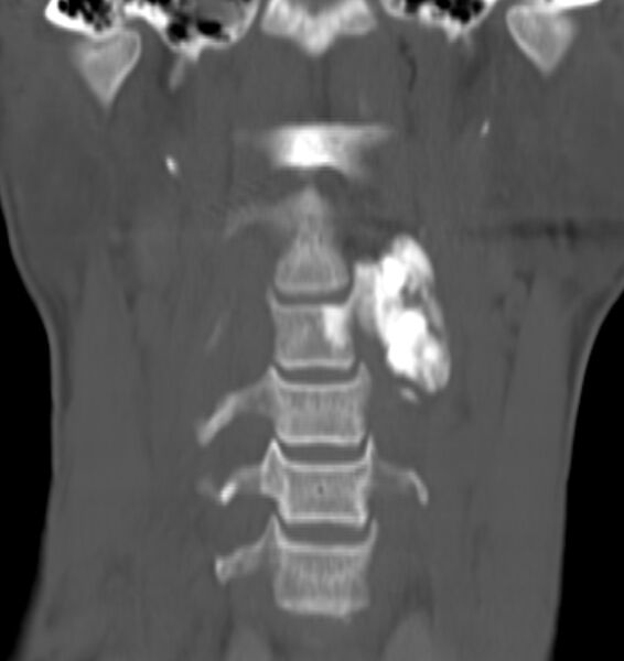
Author:Case courtesy of Dr Ammar Haouimi, Radiopaedia.org, rID: 85114 Source: https://radiopaedia.org/cases/osteoblastoma-cervical-spine?lang=us Description: CT scan neck - There is an expansile lytic/sclerotic bony mass centered on the left half of C3 vertebral body and pedicle, encasing the transverse foramen. The lytic component is mainly central. No soft tissue component is seen.
Licensing
| This work is licensed under the Creative Commons Attribution-NonCommersial-ShareAlike 4.0 License. |
File history
Click on a date/time to view the file as it appeared at that time.
| Date/Time | Thumbnail | Dimensions | User | Comment | |
|---|---|---|---|---|---|
| current | 12:07, 1 July 2021 |  | 708 × 750 (119 KB) | Whispyhistory (talk | contribs) | Author:Case courtesy of Dr Ammar Haouimi, Radiopaedia.org, rID: 85114 Source: https://radiopaedia.org/cases/osteoblastoma-cervical-spine?lang=us Description: CT scan neck - There is an expansile lytic/sclerotic bony mass centered on the left half of C3 vertebral body and pedicle, encasing the transverse foramen. The lytic component is mainly central. No soft tissue component is seen. |
You cannot overwrite this file.
File usage
The following file is a duplicate of this file (more details):
- File:Osteoblastoma - cervical spine (Radiopaedia 85114-100667 Coronal 1).jpg from a shared repository
The following page uses this file: