Halo sign
| Halo sign | |
|---|---|
| Differential diagnosis | Giant cell arteritis,[1] |
The halo sign is a medical sign used to describe findings on ultrasound in giant cell arteritis,[1][2] the ground-glass pattern around blood filled nodules and fungal infections in the lungs seen on CT scan,[3] and the double ring pattern seen on a material with bloody cerebrospinal fluid.[4][5][6]
Giant cell arteritis
A finding of a dark halo around the arterial lumen on ultrasound that suggests the diagnosis of temporal arteritis is also called a halo sign.[7] The standard diagnostic test for temporal arteritis is biopsy; however, ultrasound and MRI show promise for replacing it.[8]
Lungs
The halo sign is also understood as a region of ground-glass attenuation surrounding a pulmonary nodule on an X-ray computed tomography (CT scan) of the chest. It can be associated with hemorrhagic nodules, tumors, or inflammatory processes, but is most commonly known as an early radiographic sign of invasive pulmonary infection by the fungus species Aspergillus.[3][9]
When the central ground-glass opacity is surrounded by denser air–space consolidation in the shape of a ring or crescent, it is sometimes described as a reverse halo sign, seen in several lung conditions including fungal infections, tuberculosis, community-acquired pneumonia, Wegener granulomatosis, lymphomatoid granulomatosis and sarcoidosis.[10] It is sometimes known as the Atoll sign,[11] named for the coral atoll.[12]
Cerebrospinal fluid
The halo sign, also described as a double-ring sign, is sometimes used to determine whether bloody discharge from the nose or ears contains cerebrospinal fluid.[6] It is based on the mechanism of chromatography, where various components of a liquid mixture separate differently. It is not specific for cerebrosppinal fluid, as tears, nasal discharge and saline can also produce the sign.[4][5] To perform the test, the leaking fluid is dripped onto a 4x4 gauze or towel. Positive results are indicated by blood coalescing into the center, leaving an outer ring of cerebrospinal fluid.[6]
Deuel's halo sign
Deuel's halo sign Archived 2021-11-10 at the Wayback Machine is a sign showing the separation of the subcutaneous fat of the fetal scalp from the skull bones, seen on an medical imaging in fetal death.[13][14]
History
Deuel's halo sign in fetal death was first described by Deuel in 1947.[14] The halo sign was first described in temporal arteritis in 1995.[1] In 1996, Voloudaki et al described the halo sign in CT scanning of cryptogenic organising pneumonia.[12]
Images
-

CT of lung infarction with reverse halo sign
-

Chest CT with reversed halo sign
-
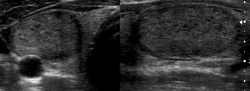
Ultrasound: halo sign of thyroid nodule


_(Radiopaedia_21891).png)
References
- ↑ 1.0 1.1 1.2 Schmidt, Wolfgang A. (23 February 2018). "Ultrasound in the diagnosis and management of giant cell arteritis". Rheumatology. 57 (2): ii22–ii31. doi:10.1093/rheumatology/kex461. Archived from the original on 14 June 2020. Retrieved 3 June 2021.
- ↑ Fernández-Fernández, Elisa; Monjo-Henry, Irene; Bonilla, Gema; Plasencia, Chamaida; Miranda-Carús, María-Eugenia; Balsa, Alejandro; De Miguel, Eugenio (1 September 2020). "False positives in the ultrasound diagnosis of giant cell arteritis: some diseases can also show the halo sign". Rheumatology. 59 (9): 2443–2447. doi:10.1093/rheumatology/kez641. ISSN 1462-0324. PMID 31953951. Archived from the original on 3 June 2021. Retrieved 1 June 2021.
- ↑ 3.0 3.1 Collins, Jannette; Stern, Eric J. (2008). "2. Signs and patterns of lung disease". Chest Radiology: The Essentials (2nd ed.). Philadelphia: Wolters Kluwer. p. 21. ISBN 978-0-7817-6314-1. Archived from the original on 2021-08-29. Retrieved 2021-06-03.
- ↑ 4.0 4.1 Sunder, Ravi; Tyler, Kevin (19 March 2013). "Basal skull fracture and the halo sign". CMAJ : Canadian Medical Association Journal. 185 (5): 416. doi:10.1503/cmaj.120055. ISSN 0820-3946. PMID 22891200. Archived from the original on 17 November 2020. Retrieved 3 June 2021.
- ↑ 5.0 5.1 Akhaddar, Ali (2017). "7. Post traumatic meningitis". Atlas of Infections in Neurosurgery and Spinal Surgery. Springer. p. 65. ISBN 978-3-319-60085-7. Archived from the original on 2021-08-28. Retrieved 2021-06-03.
- ↑ 6.0 6.1 6.2 Wilkins, Lippincott Williams & (2008). Interpreting Signs and Symptoms. Lippincott Williams & Wilkins. p. 72. ISBN 978-1-58255-668-0. Archived from the original on 2021-08-28. Retrieved 2021-06-03. Cite error: Invalid
<ref>tag; name "Wilkins2008" defined multiple times with different content - ↑ Schmidt W, Kraft H, Vorpahl K, Völker L, Gromnica-Ihle E (1997). "Color duplex ultrasonography in the diagnosis of temporal arteritis". N Engl J Med. 337 (19): 1336–42. doi:10.1056/NEJM199711063371902. PMID 9358127.
- ↑ Bley TA, Brink I, Reinhard M (April 2006). "[Imaging procedures for giant cell arteritis (Horton's disease)]". Ophthalmologe (in Deutsch). 103 (4): 308–16. doi:10.1007/s00347-006-1323-x. PMID 16538476.
- ↑ Jiang, Tao; Zhang, Yangling; Wu, Shanshan; Mao, Jujiang (2020). "4. Chest". In Gao, Bo; McKinney, Alexander M. (eds.). Classic Imaging Signs: A Guide to the Whole Body. Springer. pp. 131–132. ISBN 978-3-030-56347-9. Archived from the original on 2021-08-28. Retrieved 2021-06-01.
- ↑ Godoy, M C B; Viswanathan, C; Marchiori, E; Truong, M T; Benveniste, M F; Rossi, S; Marom, E M (September 2012). "The reversed halo sign: update and differential diagnosis". The British Journal of Radiology. 85 (1017): 1226–1235. doi:10.1259/bjr/54532316. ISSN 0007-1285. PMID 22553298. Archived from the original on 2020-12-09. Retrieved 2021-06-03.
- ↑ Chiarenza, Alessandra; Esposto Ultimo, Luca; Falsaperla, Daniele; Travali, Mario; Foti, Pietro Valerio; Torrisi, Sebastiano Emanuele; Schisano, Matteo; Mauro, Letizia Antonella; Sambataro, Gianluca; Basile, Antonio; Vancheri, Carlo; Palmucci, Stefano (4 December 2019). "Chest imaging using signs, symbols, and naturalistic images: a practical guide for radiologists and non-radiologists". Insights into Imaging. 10 (1): 114. doi:10.1186/s13244-019-0789-4. ISSN 1869-4101. PMID 31802270. Archived from the original on 14 July 2021. Retrieved 3 June 2021.
- ↑ 12.0 12.1 Han, Jason; Xiang, Hao; Ridley, William E.; Ridley, Lloyd J. (2018). "Atoll sign: High resolution CT". Journal of Medical Imaging and Radiation Oncology. 62 (S1): 17–17. doi:10.1111/1754-9485.05_12785. ISSN 1754-9485.
- ↑ Shaff MI (April 1975). "An evaluation of the radiological signs of fetal death". S. Afr. Med. J. 49 (18): 736–8. PMID 1169818.
- ↑ 14.0 14.1 Ohlson, Lars (1 January 1962). "Deuel'S Halo Sign and its Comparative Value in the Diagnosis of Foetal Death". Acta Radiologica. os-57 (1): 57–64. doi:10.1177/028418516205700109. ISSN 0001-6926. Archived from the original on 28 August 2021. Retrieved 3 June 2021.
This medical sign article is a stub. You can help MDWiki by expanding it. |