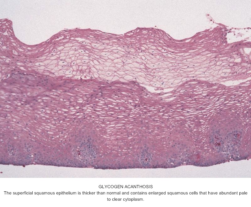
Glycogenic acanthosis
| Glycogenic acanthosis | |
|---|---|
 | |
| Endoscopic image of glycogenic acanthosis found incidentally | |
| Specialty | Gastroenterology |
Glycogenic acanthosis are small raised white plaques commonly seen in the esophageal mucosa.[1] It is seen incidentally in 3.5% of gastroscopies.[2]
Signs and symptoms
On gastroscopy, glycogenic acanthosis is seen as a multitude of small white raised plaques of 2 mm to 10 mm in size, which may be seen throughout the esophagus. They tend to occur on esophageal folds, and may be missed if the esophagus is not well distended with air. It may be seen on esophageal x-rays; it is not seen on standard esophograms, but can be seen with double-contrast studies.[3] Biopsies of the lesions show hypertrophied stratified squamous mucosa with glycogen deposition in the mucosa.[1]
Clinically, mild glycogenic acanthosis is a normal finding, and does not progress to esophageal cancer or to stricture.[4] It is not related to leukoplakia, and is not dysplastic or premalignant. It was originally thought to be associated with gastroesophageal reflux disease (GERD), but the association is not entirely clear.[2] One report also shows an association with celiac disease, but again, this has not shown been beyond that. Extensive glycogenic acanthosis has been shown to be associated with Cowden's syndrome.[5]
Diagnosis
Glycogenic acanthosis is characterized by epithelial hyperplasia, with an increased number of enlarged epithelial cells containing abundant glycogen. There is no associated hyperkeratosis, inflammation, dysplasia, or cellular atypia.[4]
References
- ^ a b Ghahremani GG, Rushovich AM. Glycogenic acanthosis of the esophagus: radiographic and pathologic features. Gastrointest Radiol. 1984;9(2):93-8. PMID 6745598.
- ^ a b Vadva MD, Triadafilopoulos G. Glycogenic acanthosis of the esophagus and gastroesophageal reflux. J Clin Gastroenterol. 1993 Jul;17(1):79-83. PMID 8409304
- ^ Glick SN, Teplich SK, Goldstein J, Stead JA, Zitomer N. Glycogenic Acanthosis of the Esophagus. Amer J Radiol 1982139:683-688
- ^ a b Pathology Outlines website: http://pathologyoutlines.com/esophagus.html#glycogenic Accessed 6 January 2009.
- ^ Kay PS, Soetikno RM, Mindelzun R, Young HS. Diffuse esophageal glycogenic acanthosis: an endoscopic marker of Cowden's disease. Am J Gastroenterol. 1997 Jun;92(6):1038-40 PMID 9177527