File:Recurrent-chondromyxoid-fibroma-of-the-humerus.jpg
Jump to navigation
Jump to search
Size of this preview: 255 × 598 pixels. Other resolution: 267 × 626 pixels.
Original file (267 × 626 pixels, file size: 12 KB, MIME type: image/jpeg)
Summary
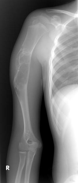
Author:Case courtesy of Dr Mohammad A. ElBeialy, Radiopaedia.org, rID: 26416 Source:https://radiopaedia.org/cases/recurrent-chondromyxoid-fibroma-of-the-humerus?lang=gb Description:X-ray right upper arm - A well-defined right proximal humeral diaphyseal expansile, multilocular lesion with a geographic, sclerotic margin. No matrix calcification and no visible periosteal reaction.
Licensing
| This work is licensed under the Creative Commons Attribution-NonCommersial-ShareAlike 4.0 License. |
File history
Click on a date/time to view the file as it appeared at that time.
| Date/Time | Thumbnail | Dimensions | User | Comment | |
|---|---|---|---|---|---|
| current | 16:12, 5 July 2021 | | 267 × 626 (12 KB) | Whispyhistory (talk | contribs) | Author:Case courtesy of Dr Mohammad A. ElBeialy, Radiopaedia.org, rID: 26416 Source:https://radiopaedia.org/cases/recurrent-chondromyxoid-fibroma-of-the-humerus?lang=gb Description:X-ray right upper arm - A well-defined right proximal humeral diaphyseal expansile, multilocular lesion with a geographic, sclerotic margin. No matrix calcification and no visible periosteal reaction. |
You cannot overwrite this file.
File usage
There are no pages that use this file.