No higher resolution available.
This file is from a shared repository and may be used by other projects.
The description on its file description page there is shown below.
License
Attribution-NonCommercial-ShareAlike 3.0 Unported (CC BY-NC-SA 3.0)]
Summary
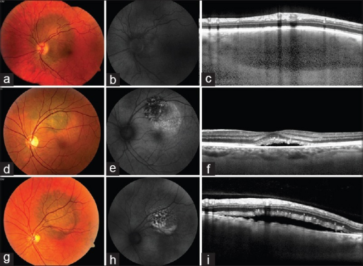
Author:Rishi P, Koundanya VV, Shields CL ,Shri Bhagwan Mahavir Vitreoretinal Services, Sankara Nethralaya (Openi/National Library of medicine) Source:https://openi.nlm.nih.gov/detailedresult?img=PMC4399118_IJO-63-110-g001&query=Uveal%20melanoma&it=xg&req=4&npos=60 Description:F1: Clinical and imaging features of choroidal melanocytic lesions. Case 1: Circumpapillary pigmented choroidal lesion lacking orange pigment (a). Fundus autoflouroscense (b) is indistinct with no lipofuscin. Enhanced depth imaging-optical coherence tomography (c) normal photoreceptors and absence of SRF, consistent with choroidal nevus. Case 2: Pigmented choroidal lesion with overlying orange pigment, diffuse hyperautofluorescence (e) and the subfoveal fluid with shaggy photoreceptors (f) suggestive of choroidal melanoma. Case 3: Pigmented choroidal lesion with orange pigment (g), patchy hyperautofluorescence with sedimentation (h), and overlying SRF and shaggy photoreceptors (i), suggestive of choroidal melanoma
File history
Click on a date/time to view the file as it appeared at that time.
| Date/Time | Thumbnail | Dimensions | User | Comment |
|---|
| current | 18:56, 7 November 2021 |  | 512 × 375 (281 KB) | Ozzie10aaaa | Author:Rishi P, Koundanya VV, Shields CL ,Shri Bhagwan Mahavir Vitreoretinal Services, Sankara Nethralaya (Openi/National Library of medicine) Source:https://openi.nlm.nih.gov/detailedresult?img=PMC4399118_IJO-63-110-g001&query=Uveal%20melanoma&it=xg&req=4&npos=60 Description:F1: Clinical and imaging features of choroidal melanocytic lesions. Case 1: Circumpapillary pigmented choroidal lesion lacking orange pigment (a). Fundus autoflouroscense (b) is indistinct with no lipofuscin. Enhanced de... |
File usage
The following page uses this file: