File:PMC3743519 idr-6-079Fig1.png
Jump to navigation
Jump to search
No higher resolution available.
PMC3743519_idr-6-079Fig1.png (512 × 456 pixels, file size: 184 KB, MIME type: image/png)
File history
Click on a date/time to view the file as it appeared at that time.
| Date/Time | Thumbnail | Dimensions | User | Comment | |
|---|---|---|---|---|---|
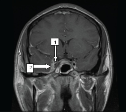
| current | 22:39, 13 October 2021 | | 512 × 456 (184 KB) | Ozzie10aaaa | Author:Clarke M, Enuh H, Saverimuttu J, Nfonoyim J ,Department of Medicine, Richmond University Medical Center (Openi/National Library of National) Source:https://openi.nlm.nih.gov/detailedresult?img=PMC3743519_idr-6-079Fig1&query=Cavernous%20sinus%20thrombosis&it=xg&req=4&npos=3 Description:f1-idr-6-079: A band of enhancement measuring 6 mm in thickness is seen along the medial margin of the right temporal convexity (arrow #2). Increased T2 signal in the region of the right cavernous sinus w... |
File usage
The following page uses this file: