File:PMC3534218 2045-3329-2-22-3.png
Jump to navigation
Jump to search
No higher resolution available.
PMC3534218_2045-3329-2-22-3.png (512 × 314 pixels, file size: 119 KB, MIME type: image/png)
File history
Click on a date/time to view the file as it appeared at that time.
| Date/Time | Thumbnail | Dimensions | User | Comment | |
|---|---|---|---|---|---|
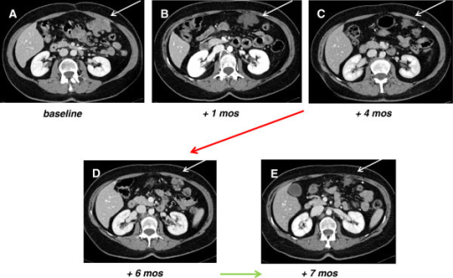
| current | 22:26, 15 November 2021 | | 512 × 314 (119 KB) | Ozzie10aaaa | Author:Stacchiotti S, Dagrada GP, Morosi C, Negri T, Romanini A, Pilotti S, Gronchi A, Casali PG ,Department of Cancer Medicine, Adult Sarcoma Medical Oncology Unit, Fondazione IRCCS Istituto Nazionale Tumori Milan (Openi/National Library of Medicine) Source:https://openi.nlm.nih.gov/detailedresult?img=PMC3534218_2045-3329-2-22-3&query=sunitinib&it=xg&req=4&npos=34 Description:F3: Computed Tomography scan (CT) tumor assessment. In panels A, B, C (white arrows) CT, venous phase, after contrast... |
File usage
The following page uses this file: