File:PMC3380688 jovr-5-2-90-642-2-pbf1.png
Jump to navigation
Jump to search
PMC3380688_jovr-5-2-90-642-2-pbf1.png (512 × 174 pixels, file size: 112 KB, MIME type: image/png)
Summary
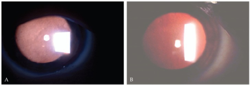
Author:Javadi MA, Rezaei-Kanavi M, Javadi A, Naghshgar N,Ophthalmic Research Center, Shahid Beheshti University of Medical Sciences, Tehran, Iran.(Openi National Library of Medicine) Source:https://openi.nlm.nih.gov/detailedresult?img=PMC3380688_jovr-5-2-90-642-2-pbf1&query=Meesmann%20corneal%20dystrophy&it=xg&req=4&npos=18 Description:f1-jovr-5-2-90-642-2-pb: Diffuse intraepithelial microcystic lesions on slitlamp biomicroscopy visible by retroillumination in the first (A) and second (B) patient.
Licensing
File history
Click on a date/time to view the file as it appeared at that time.
| Date/Time | Thumbnail | Dimensions | User | Comment | |
|---|---|---|---|---|---|
| current | 16:31, 29 July 2021 | 512 × 174 (112 KB) | Ozzie10aaaa (talk | contribs) | Author:Javadi MA, Rezaei-Kanavi M, Javadi A, Naghshgar N,Ophthalmic Research Center, Shahid Beheshti University of Medical Sciences, Tehran, Iran.(Openi National Library of Medicine) Source:https://openi.nlm.nih.gov/detailedresult?img=PMC3380688_jovr-5-2-90-642-2-pbf1&query=Meesmann%20corneal%20dystrophy&it=xg&req=4&npos=18 Description:f1-jovr-5-2-90-642-2-pb: Diffuse intraepithelial microcystic lesions on slitlamp biomicroscopy visible by retroillumination in the first (A) and second (B) pat... |
You cannot overwrite this file.
File usage
The following page uses this file:
