File:PMC2519249 ehn29605.png
Jump to navigation
Jump to search
No higher resolution available.
PMC2519249_ehn29605.png (400 × 251 pixels, file size: 80 KB, MIME type: image/png)
File history
Click on a date/time to view the file as it appeared at that time.
| Date/Time | Thumbnail | Dimensions | User | Comment | |
|---|---|---|---|---|---|
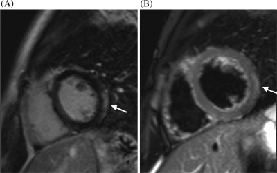
| current | 20:51, 2 January 2022 | | 400 × 251 (80 KB) | Ozzie10aaaa | Author:Dennert R, Crijns HJ, Heymans S, Department of Cardiology, CARIM, University Hospital(Openi/National Library of Medicine) Source:https://openi.nlm.nih.gov/detailedresult?img=PMC2519249_ehn29605&query=Autoimmune%20myocarditis&it=xg&req=4&npos=70 Description:EHN296F5: Cardiovascular magnetic resonance image. Short-axis cardiac magnetic resonance imaging of a patient with acute myocarditis (A) T2-weighted image, showing regional oedema of the lateral left ventricle predominantly subepicar... |
File usage
The following page uses this file: