File:Osteofibrous-dysplasia-of-the-tibia.jpeg
Jump to navigation
Jump to search

Size of this preview: 444 × 600 pixels. Other resolutions: 178 × 240 pixels | 355 × 480 pixels | 568 × 768 pixels | 758 × 1,024 pixels | 2,416 × 3,264 pixels.
Original file (2,416 × 3,264 pixels, file size: 737 KB, MIME type: image/jpeg)
Summary
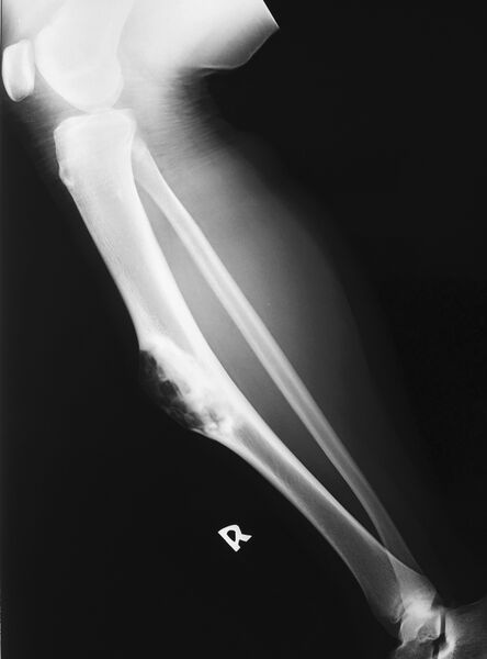
Author: Case courtesy of Dr Ian Bickle, Radiopaedia.org, rID: 41652
Source:https://radiopaedia.org/cases/osteofibrous-dysplasia-of-the-tibia?lang=us
Description: Plain X-ray of tibia - Expansile cortical based lucent lesion with a narrow zone of transition in the mid tibial diaphysis. The anterior cortext is thinned, but not breeched. Internal 'soap bubble'appearance to the lesion which extends into the medulla and has a sclerotic rim.
Licensing
| This work is licensed under the Creative Commons Attribution-NonCommersial-ShareAlike 4.0 License. |
File history
Click on a date/time to view the file as it appeared at that time.
| Date/Time | Thumbnail | Dimensions | User | Comment | |
|---|---|---|---|---|---|
| current | 18:12, 12 May 2021 | | 2,416 × 3,264 (737 KB) | Whispyhistory (talk | contribs) | Author: Case courtesy of Dr Ian Bickle, Radiopaedia.org, rID: 41652 Source:https://radiopaedia.org/cases/osteofibrous-dysplasia-of-the-tibia?lang=us Description: Plain X-ray of tibia - Expansile cortical based lucent lesion with a narrow zone of transition in the mid tibial diaphysis. The anterior cortext is thinned, but not breeched. Internal 'soap bubble'appearance to the lesion which extends into the medulla and has a sclerotic rim. |
You cannot overwrite this file.
File usage
There are no pages that use this file.