File:Non-ossifying-fibroma-6-2.jpg
Jump to navigation
Jump to search

Size of this preview: 608 × 600 pixels. Other resolutions: 243 × 240 pixels | 486 × 480 pixels | 778 × 768 pixels | 1,038 × 1,024 pixels | 1,961 × 1,935 pixels.
Original file (1,961 × 1,935 pixels, file size: 191 KB, MIME type: image/jpeg)
Summary
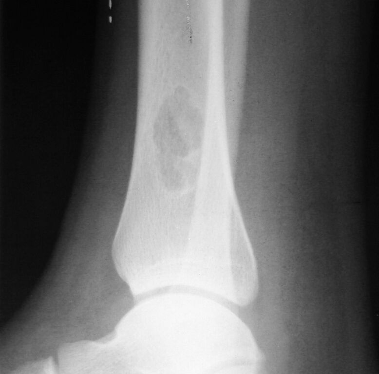
Author: Case courtesy of Dr Mohammad Osama Hussein Yonso, Radiopaedia.org, rID: 17804
Source: https://radiopaedia.org/cases/non-ossifying-fibroma-6?lang=us
Description: Lateral view plain X-ray: Non-ossifying fibroma of the distal tibia: well-defined outline of the tumor with sharply defined sclerotic margins.
Licensing
| This work is licensed under the Creative Commons Attribution-NonCommersial-ShareAlike 4.0 License. |
File history
Click on a date/time to view the file as it appeared at that time.
| Date/Time | Thumbnail | Dimensions | User | Comment | |
|---|---|---|---|---|---|
| current | 10:34, 11 May 2021 | | 1,961 × 1,935 (191 KB) | Whispyhistory (talk | contribs) | Author: Case courtesy of Dr Mohammad Osama Hussein Yonso, Radiopaedia.org, rID: 17804 Source: https://radiopaedia.org/cases/non-ossifying-fibroma-6?lang=us Description: Lateral view plain X-ray: Non-ossifying fibroma of the distal tibia: well-defined outline of the tumor with sharply defined sclerotic margins. |
You cannot overwrite this file.
File usage
The following file is a duplicate of this file (more details):
- File:Non-ossifying fibroma (Radiopaedia 17804-17565 Lateral 1).jpg from a shared repository
.jpg)
There are no pages that use this file.