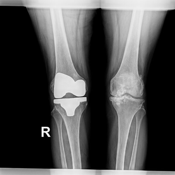
File:Baker cyst with synovial chondromatosis (Radiopaedia 69206-78994 Frontal 1).jpg

From WikiProjectMed
Jump to navigation Jump to search

Original file(4,298 × 4,300 pixels, file size: 723 KB, MIME type: image/jpeg)

This file is from a shared repository and may be used by other projects. The description on its file description page there is shown below.

Summary:

Description
  • Radiopaedia case ID: 69206
  • Image ID: 49946459
  • Plane projection: Frontal
  • Modality: X-ray
  • System: Musculoskeletal
  • Findings: Left knee joint shows triple compartment osteoarthritis seen as decreased medial femorotibial compartment joint space, subchondral sclerosis, medial compartment articular cartilage thinning, marginal and patellofemoral osteophyte formation. Multiple globular calcific lesions in Baker's cyst is in keeping with secondary synovial chondromatosis. Total knee arthroplasty is seen on the right side. Chondromalacia patellae as subchondral sclerosis and cyst formation are seen at the medial and lateral facet joints of patella bilaterally.
Date Published: 3rd Jul 2019
Source https://radiopaedia.org/cases/baker-cyst-with-synovial-chondromatosis
Author Bahman Rasuli
Permission
(Permission-reusing-text)
http://creativecommons.org/licenses/by-nc-sa/3.0/

Licensing:

Attribution-NonCommercial-ShareAlike 3.0 Unported (CC BY-NC-SA 3.0)

File history

Click on a date/time to view the file as it appeared at that time.

Date/TimeThumbnailDimensionsUserComment
current23:56, 5 June 2021Thumbnail for version as of 23:56, 5 June 20214,298 × 4,300 (723 KB)Radiopaedia project rID:69206 (batch #3577-1 A1)

The following 2 pages use this file: