Connecting stalk
| Connecting stalk | |
|---|---|
 Diagram showing the expansion of amnion and delimitation of the umbilical cord | |
 Section through the embryo | |
| Details | |
| Precursor | Extraembryonic mesoderm |
| Identifiers | |
| Latin | pedunculus connectans |
| TE | stalk_by_E5.11.3.1.1.0.4 E5.11.3.1.1.0.4 |
| Anatomical terminology | |
The connecting stalk, or body stalk, is an embryonic structure that is formed by the third week of development and connects the embryo to its shell of trophoblasts. The connecting stalk is derived from the extraembryonic mesoderm.[1] Initially it lies caudally to the trilaminar germ disc, but, with subsequent embryonic folding, the body stalk assume a more ventral position.[2] Progressive expansion of the amnion from the umbilical ring (surrounding the roots of the vitelline duct and connecting stalk) creates a tube with a covering of amniotic membrane with allantois and umbilical vessels as its content and mesoderm of the connecting stalk as the ground substance. This extraembryonic mesodermal ground substance forms the future Wharton's jelly.[2] The amniotic membrane and its contents form the umbilical cord that connects the embryo and the placenta.[3][4]
The root of the connecting stalk contains the allantois as a diverticulum of hindgut endoderm along with umbilical vessels.[5][2]
Anomalies are usually referred to as body stalk anomalies and occur in approximately 1 in 15,000 births.[6] They are due to defects in the formation of the cephalic, caudal, and lateral embryonic body folds,[7] that result in a reduced or absent umbilical cord.[8]
Additional images
-
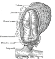 Human embryo—length, 2 mm. Dorsal view, with the amnion laid open. X 30.
 -
 Human embryo of 2.6 mm.
 -
 Model of human embryo 1.3 mm. long.
 -
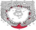 Section through ovum imbedded in the uterine decidua.
 -
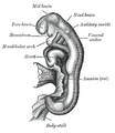 Embryo between eighteen and twenty-one days.


References
- ^ Schoenwolf, Gary C.; Bleyl, Steven B.; Brauer, Philip R.; Francis-West, Philippa H. (2012-05-07). Larsen's Human Embryology: with STUDENT CONSULT Online Access. Elsevier Health Sciences. p. 57. ISBN 9781455727919.
- ^ a b c Heil, Jenna; Bordoni, Bruno (2022-04-21). "Embryology, Umbilical Cord". StatPearls. PMID 32491422.
- ^ Sadler, T. W. (2010). Langman's medical embryology (11th ed.). Philadelphia: Lippincott William & Wilkins. pp. 64–65. ISBN 9780781790697.
- ^ Larsen's Embryology, 5th edition, p138.
- ^ Larsen, William J. (2001). Human embryology (3rd ed.). New York: Churchill Livingstone. p. 138. ISBN 0443065837.
- ^ Asim Kurjak (30 June 2013). Donald School Textbook of Transvaginal Sonography. JP Medical Ltd. p. 28. ISBN 978-93-5090-473-2.
- ^ Diana W. Bianchi; Timothy M. Crombleholme; Mary E. D'Alton (1 January 2000). Fetology: Diagnosis & Management of the Fetal Patient. McGraw Hill Professional. ISBN 978-0-8385-2570-8.
- ^ Kocherla, K; Kumari, V; Kocherla, PR (January 2015). "Prenatal diagnosis of body stalk complex: A rare entity and review of literature". The Indian Journal of Radiology & Imaging. 25 (1): 67–70. doi:10.4103/0971-3026.150162. PMC 4329692. PMID 25709170.
External links
- Swiss embryology (from UL, UB, and UF) hdisqueembry/triderm0
This developmental biology article is a stub. You can help Wikipedia by expanding it. |