AT-hook
| AT-hook | |||||||||
|---|---|---|---|---|---|---|---|---|---|
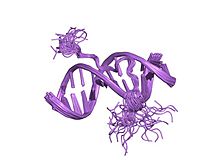
 solution structure of a complex of the second dna binding domain of human hmg-i(y) bound to dna dodecamer containing the prdii site of the interferon-beta promoter, nmr, 35 structures | |||||||||
| Identifiers | |||||||||
| Symbol | AT_hook | ||||||||
| Pfam | PF02178 | ||||||||
| InterPro | IPR017956 | ||||||||
| SMART | AT_hook | ||||||||
| SCOP2 | 2eze / SCOPe / SUPFAM | ||||||||
| |||||||||

The AT-hook is a DNA-binding motif present in many proteins, including the high mobility group (HMG) proteins,[1] DNA-binding proteins from plants[2] and hBRG1 protein, a central ATPase of the human switching/sucrose non-fermenting (SWI/SNF) remodeling complex.[3]
Structure
This motif consists of a conserved, palindromic, core sequence of proline-arginine-glycine-arginine-proline, although some AT-hooks contain only a single proline in the core sequence. AT-hooks also include a variable number of positively charged lysine and arginine residues on either side of the core sequence.[4] The AT-hook binds to the minor groove of adenine-thymine (AT) rich DNA, hence the AT in the name. The rest of the name derives from a predicted asparagine/aspartate "hook" in the earliest AT-hooks reported in 1990.[5] In 1997 structural studies using NMR determined that a DNA-bound AT-hook adopted a crescent or hook shape around the minor groove of a target DNA strand (pictured at right).[6] HMGA proteins contain three AT-hooks, although some proteins contain as many as 30.[5] The optimal binding sequences for AT-hook proteins are repeats of the form (ATAA)n or (TATT)n, although the optimal binding sequences for the core sequence of the AT-hook are AAAT and AATT.[7]
The DNA dodecamer has eight consecutive AT base pairs, allowing the AT-hook to be positioned in several positions, with the preferred position being at one of the AATT regions to fully occupy the minor groove. Van der Waals interactions of the AT-hook with the adenines play an important role for the specificity of the position.[8] Van der Waals interactions of the AT-hook with the adenines play an important role for the specificity of the position.[8]


The figure shows the position of the main chain to allow hydrogen bonds with the minor groove thymine oxygen atoms. The interactions shown, caused the DNA to bend, extending the minor groove. The distorted DNA causes the complementary major groove to form interactions between the side chains.
Function
AT-hook proteins can form hydrogen bonds between NH groups of Gly37 and Arg38 on the main-chain and thymine oxygen atoms in the minor groove, which bends the DNA and widens the minor groove.[8] The binding to the minor groove facilitates binding of other proteins in the major groove.[9] That enables HMG proteins to regular expression of genes and influence biological processes.
The AT-hooks have also been proposed to anchor chromatin-modifying proteins to AT-rich DNA sequences through their association with chromatin remodeling, histone modifications, and chromatin insulator function.[9]
Clinical significance
Alterations or abnormal expression of the HMG proteins have led to metabolic disorders, such as obesity, type 2 diabetes, and cancer.[8]
References
- ^ Reeves R, Beckerbauer L (May 2001). "HMGI/Y proteins: flexible regulators of transcription and chromatin structure". Biochimica et Biophysica Acta (BBA) - Gene Structure and Expression. 1519 (1–2): 13–29. doi:10.1016/S0167-4781(01)00215-9. PMID 11406267.
- ^ Meijer AH, van Dijk EL, Hoge JH (June 1996). "Novel members of a family of AT hook-containing DNA-binding proteins from rice are identified through their in vitro interaction with consensus target sites of plant and animal homeodomain proteins". Plant Molecular Biology. 31 (3): 607–618. doi:10.1007/BF00042233. PMID 8790293. S2CID 24687309.
- ^ Singh M, D'Silva L, Holak TA (2006). "DNA-binding properties of the recombinant high-mobility-group-like AT-hook-containing region from human BRG1 protein". Biological Chemistry. 387 (10–11): 1469–1478. doi:10.1515/BC.2006.184. PMID 17081121. S2CID 26580880.
- ^ Reeves R (October 2001). "Molecular biology of HMGA proteins: hubs of nuclear function". Gene. 277 (1–2): 63–81. doi:10.1016/S0378-1119(01)00689-8. PMID 11602345.
- ^ a b Reeves R, Nissen MS (May 1990). "The A.T-DNA-binding domain of mammalian high mobility group I chromosomal proteins. A novel peptide motif for recognizing DNA structure". The Journal of Biological Chemistry. 265 (15): 8573–8582. doi:10.1016/S0021-9258(19)38926-4. PMID 1692833.
- ^ Huth JR, Bewley CA, Nissen MS, Evans JN, Reeves R, Gronenborn AM, Clore GM (August 1997). "The solution structure of an HMG-I(Y)-DNA complex defines a new architectural minor groove binding motif". Nature Structural Biology. 4 (8): 657–665. doi:10.1038/nsb0897-657. PMID 9253416. S2CID 2183841.
- ^ Reeves R (October 2000). "Structure and function of the HMGI(Y) family of architectural transcription factors". Environmental Health Perspectives. 108 Suppl 5 (Suppl 5): 803–809. doi:10.2307/3454310. JSTOR 3454310. PMID 11035986.
- ^ a b c d Fonfría-Subirós E, Acosta-Reyes F, Saperas N, Pous J, Subirana JA, Campos JL (2012). "Crystal structure of a complex of DNA with one AT-hook of HMGA1". PLOS ONE. 7 (5): e37120. Bibcode:2012PLoSO...737120F. doi:10.1371/journal.pone.0037120. PMC 3353895. PMID 22615915.
- ^ a b Filarsky M, Zillner K, Araya I, Villar-Garea A, Merkl R, Längst G, Németh A (2015). "The extended AT-hook is a novel RNA binding motif". RNA Biology. 12 (8): 864–876. doi:10.1080/15476286.2015.1060394. PMC 4615771. PMID 26156556.