File:Two-pore domain potassium channel K2P1 PDB-3ukm.png
Jump to navigation
Jump to search
Size of this preview: 510 × 600 pixels. Other resolutions: 204 × 240 pixels | 408 × 480 pixels | 850 × 1,000 pixels.
Original file (850 × 1,000 pixels, file size: 438 KB, MIME type: image/png)
Summary
| Description |
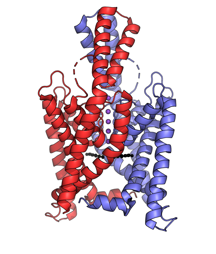
English: Homodimeric two-pore domain potassium channel K2P1 with bound potassium ions (purple). Four helices extend away from the membrane, into the extra-cellular space. Voids in the channel protein are filled with electron density consistent with alkyl chains (black). PDB: 3UKM |
| Date |
(upload to Wikimedia Commons), 9 November 2011 (deposition at PDB) |
| Source | Atom coordinates: https://www.rcsb.org/structure/3UKM; Visualization: Own work |
| Author |
Deposition authors: Long, S.B., Miller, A.N.; Visualization author: Synpath |
Licensing
I, the copyright holder of this work, hereby publish it under the following license:
| This file is made available under the Creative Commons CC0 1.0 Universal Public Domain Dedication. | |
| The person who associated a work with this deed has dedicated the work to the public domain by waiving all of their rights to the work worldwide under copyright law, including all related and neighboring rights, to the extent allowed by law. You can copy, modify, distribute and perform the work, even for commercial purposes, all without asking permission.
|
File history
Click on a date/time to view the file as it appeared at that time.
| Date/Time | Thumbnail | Dimensions | User | Comment | |
|---|---|---|---|---|---|
| current | 00:54, 20 November 2023 | | 850 × 1,000 (438 KB) | commons>Synpath | Uploaded own work with UploadWizard |
File usage
There are no pages that use this file.