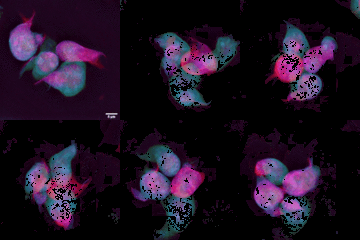
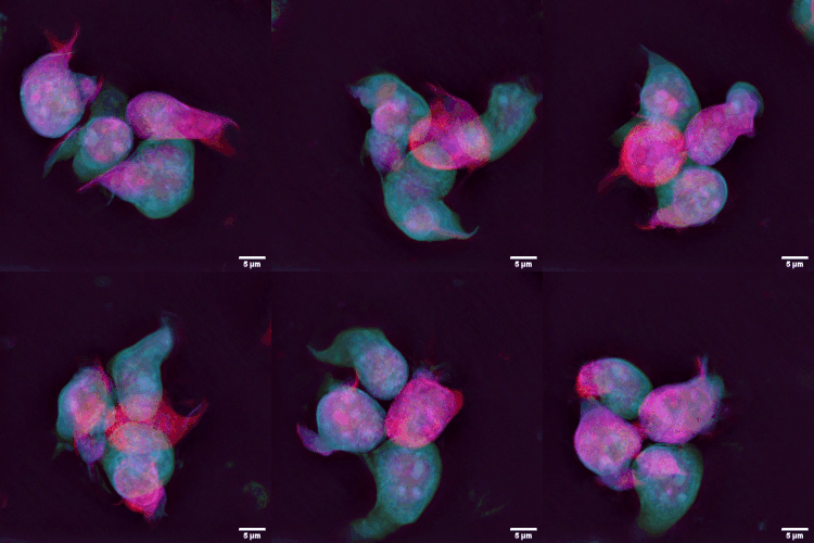
File:T cell nuclear dynamics.gif
Jump to navigation
Jump to search
T_cell_nuclear_dynamics.gif (250 × 250 pixels, file size: 3.15 MB, MIME type: image/gif, looped, 98 frames, 9.8 s)
File history
Click on a date/time to view the file as it appeared at that time.
| Date/Time | Thumbnail | Dimensions | User | Comment | |
|---|---|---|---|---|---|
| current | 17:08, 7 November 2021 |  | 250 × 250 (3.15 MB) | Ozzie10aaaa | Author:Evilonan Source:https://commons.wikimedia.org/wiki/File:T_cell_nuclear_dynamics.gif#cite_note-1 Description:English: An example of 4D live imaging of T cell nuclear dynamics imaged with a holotomographic microscope. [1] The color code was digitally post added and allows for a depth interpretation: cells closer to the wellplate surface are colored blue, while the ones further away from it are seen as pink/red. |
File usage
The following file is a duplicate of this file (more details):
- File:T cell nuclear dynamics.gif from a shared repository
There are no pages that use this file.
