File:Polymicrogyria arrows.JPG
Jump to navigation
Jump to search
No higher resolution available.
Polymicrogyria_arrows.JPG (610 × 598 pixels, file size: 80 KB, MIME type: image/jpeg)
| Description |
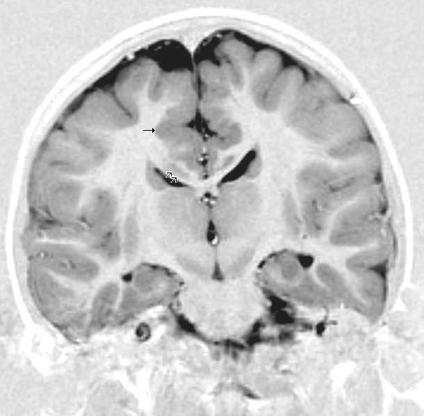
English: This child presented with seizures. The coronal true inversion recovery sequence shows thickened and disordered cortex in superior frontal and cingulate gyri bilaterally (arrow). There are small convolutions visible at the corticomedullary junction. The appearance is that of cortical dysplasia, with polymicrogyria more likely than pachygyria due to the small convolutions visible. There are also small foci of grey matter signal in the corpus callosum, deep to the dysplastic cortex (double arrows). These probably represent areas of grey matter heterotopia. |
| Date | 5 March 2008 (upload date) |
| Source | radpod.org |
| Author | Dr Laughlin Dawes |
| Permission (Reusing this file) |
author kindly mailed me permission to use this and other images on cc-by-3.0 license |
This file is licensed under the Creative Commons Attribution 3.0 Unported license.
- You are free:
- to share – to copy, distribute and transmit the work
- to remix – to adapt the work
- Under the following conditions:
- attribution – You must give appropriate credit, provide a link to the license, and indicate if changes were made. You may do so in any reasonable manner, but not in any way that suggests the licensor endorses you or your use.
File history
Click on a date/time to view the file as it appeared at that time.
| Date/Time | Thumbnail | Dimensions | User | Comment | |
|---|---|---|---|---|---|
| current | 15:47, 5 March 2008 | | 610 × 598 (80 KB) | commons>Filip em | {{Information |Description=This child presented with seizures. The coronal true inversion recovery sequence shows thickened and disordered cortex in superior frontal and cingulate gyri bilaterally (arrow). There are small convolutions visible at the corti |
File usage
The following file is a duplicate of this file (more details):
- File:Grey matter heterotopia (Radiopaedia 8234).jpg from a shared repository
.jpg)
The following page uses this file: