File:PMC5400418 rjx045f05.png
Jump to navigation
Jump to search
No higher resolution available.
PMC5400418_rjx045f05.png (512 × 512 pixels, file size: 148 KB, MIME type: image/png)
File history
Click on a date/time to view the file as it appeared at that time.
| Date/Time | Thumbnail | Dimensions | User | Comment | |
|---|---|---|---|---|---|
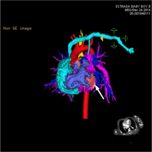
| current | 19:02, 22 October 2021 | | 512 × 512 (148 KB) | Ozzie10aaaa | Author:Pooja H. Patel, Joel Hayden, and Randy Richardson,Department of Radiology, St. Joseph's Hospital and Medical Center and Creighton University School of Medicine (Openi/National Library of medicine) Source:https://openi.nlm.nih.gov/detailedresult?img=PMC5400418_rjx045f05&query=Ivemark%20syndrome&it=xg&req=4&npos=9 Description:rjx045F5: CT 3D showing anterior heart and circulation. The left-sided subclavian vein (light blue) brings blood to the right atrium (light blue). The right ventric... |
File usage
The following page uses this file: