No higher resolution available.
This file is from a shared repository and may be used by other projects.
The description on its file description page there is shown below.
License
Attribution 4.0 International (CC BY 4.0)
Summary
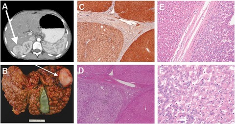
Author:Patrick R. Blackburn, Raymond D. Hickey, Rebecca A. Nace, Nasra H. Giama, Daniel L. Kraft, Andrew J. Bordner, Roongruedee Chaiteerakij,Jennifer B. McCormick, Maja Radulovic, Rondell P. Graham, 1 Michael S. Torbenson, Silvia Tortorelli, C. Ronald Scott, 4Noralane M. Lindor, Dawn S. Milliner, Devin Oglesbee, Wafa'a Al‐Qabandi, Markus Grompe, Dimitar K. Gavrilov, Mounif El‐Youssef, Karl J. Clark, 1Paldeep S. Atwal, Lewis R. Roberts, Eric W. Klee, and Stephen C.Center for Individualized Medicine, Mayo Clinic,Palo Alto Medical Foundation,Chulalongkorn University and King Chulalongkorn Memorial Hospital,Department of Pediatrics, Division of Genetic Medicine, University of Washington,5Department of Health Science Research, Mayo Clinic,Deptartment of Pediatrics, Faculty of Medicine, University of Kuwait, Department of Pediatrics, Papé Family Pediatric Research Institute (Openi/National Library of Medicine) Source:https://openi.nlm.nih.gov/detailedresult?img=PMC5108417_HUMU-37-1097-g002&query=Tyrosinemia%20type%20II&it=xg&req=4&npos=4 Description:humu23047-fig-0002: Imaging, pathological, and immunohistological study findings on the proband. A: CT scan showing the 4.8 × 3.6 × 3.6 cm3 enhancing mass in segment VI of the proband's liver. Biopsy showed moderately differentiated hepatocellular carcinoma. B: Explanted liver from the proband showing extensive macronodular cirrhosis. The arrow points to the hepatocellular carcinoma lesion shown in the CT scan in panel A. C: Fumarylacetoacetate hydrolase immunohistochemistry of liver tissue derived from the proband (10×). Macronodules were immunopositive for fumarylacetoacetate hydrolase with some variability in staining. D: Hematoxylin and eosin stain of liver tissue taken from the proband (10×). The nonneoplastic liver parenchyma showed inactive cirrhosis (stage 4) that was morphologically cryptogenic. E: Hematoxylin and eosin stained section at the tumor margin (20×). F: Hematoxylin and eosin stained section showing multiple changes in clear cell foci and mild dysplasia (40×). Pale bodies were also present.
File history
Click on a date/time to view the file as it appeared at that time.
| Date/Time | Thumbnail | Dimensions | User | Comment |
|---|
| current | 15:03, 26 October 2021 |  | 480 × 253 (290 KB) | Ozzie10aaaa | Author:Patrick R. Blackburn, Raymond D. Hickey, Rebecca A. Nace, Nasra H. Giama, Daniel L. Kraft, Andrew J. Bordner, Roongruedee Chaiteerakij,Jennifer B. McCormick, Maja Radulovic, Rondell P. Graham, 1 Michael S. Torbenson, Silvia Tortorelli, C. Ronald Scott, 4Noralane M. Lindor, Dawn S. Milliner, Devin Oglesbee, Wafa'a Al‐Qabandi, Markus Grompe, Dimitar K. Gavrilov, Mounif El‐Youssef, Karl J. Clark, 1Paldeep S. Atwal, Lewis R. Roberts, Eric W. Klee, and Stephen C.Center for... |
File usage
There are no pages that use this file.