No higher resolution available.
This file is from a shared repository and may be used by other projects.
The description on its file description page there is shown below.
License
Attribution 4.0 International (CC BY 4.0)
- &
CC0 1.0 Universal (CC0 1.0) Public Domain Dedication
Summary
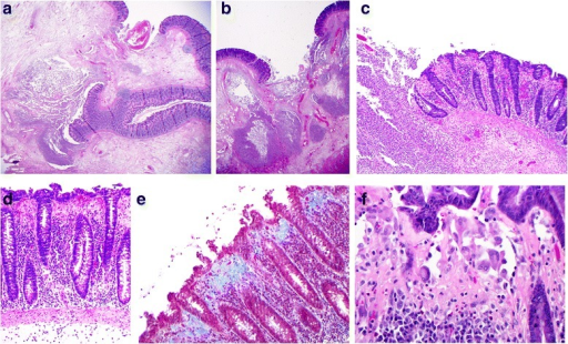
Author:Andrew Mitchell and Alexandre Dugas ,Department of Anatomic Pathology and Cytology, Maisonneuve-Rosemont Hospital, Department of Radiology, Maisonneuve-Rosemont Hospital(Openi/National Library of Medicine)Source:https://openi.nlm.nih.gov/detailedresult?img=PMC5052946_12876_2016_533_Fig3_HTML&query=Collagenous%20colitis&it=xg&req=4&npos=2 Description:Fig3: Microscopy of the resected colon: a and b Low power views (×12.5) of ulcerated bowel with abscess formation and transmural necrosis. Even at this low magnification the non-ulcerated mucosa shows clear evidence of collagenous colitis. c Medium power view (×100) of mucosal ulceration (left) and severe collagenous colitis (right). d High power view (×200) of the mucosa showing the features of collagenous colitis: a markedly thickened subepithelial collagen layer measuring up to 100 μm, surface epithelial flattening with loss of goblet cells, separation of the epithelium from the lamina propria, increased intraepithelial and lamina propria chronic inflammatory cells. e High power view (×200), Masson's trichrome stain, in which the thickened subepithelial collagen layer is stained blue. f Very high power view (×400) of multinucleated histiocytes
File history
Click on a date/time to view the file as it appeared at that time.
| Date/Time | Thumbnail | Dimensions | User | Comment |
|---|
| current | 19:26, 6 January 2022 |  | 512 × 310 (382 KB) | Ozzie10aaaa | Author:Andrew Mitchell and Alexandre Dugas ,Department of Anatomic Pathology and Cytology, Maisonneuve-Rosemont Hospital, Department of Radiology, Maisonneuve-Rosemont Hospital(Openi/National Library of Medicine)Source:https://openi.nlm.nih.gov/detailedresult?img=PMC5052946_12876_2016_533_Fig3_HTML&query=Collagenous%20colitis&it=xg&req=4&npos=2 Description:Fig3: Microscopy of the resected colon: a and b Low power views (×12.5) of ulcerated bowel with abscess formation and transmural necrosis.... |
File usage
The following page uses this file: