
No higher resolution available.
This file is from a shared repository and may be used by other projects.
The description on its file description page there is shown below.
License
Attribution 4.0 International (CC BY 4.0)
Summary
Author:Claire E L Smith, James A Poulter, Alex V Levin, Jenina E Capasso, Susan Price, Tamar Ben-Yosef, Reuven Sharony, William G Newman, Roger C Shore, Steven J Brookes, Alan J Mighell, and Chris F Inglehearn Leeds Institute of Biomedical and Clinical Sciences, St. James's University Hospital, University of Leeds,Sidney Kimmel Medical College at Thomas Jefferson University,Children's Hospital of the King's Daughters, Pediatric Ophthalmology and Ocular Genetics, Department of Clinical Genetics, Northampton General Hospital, NHS Trust Rappaport Faculty of Medicine, Technion, Haifa,The Genetic Institute and Obstetrics and Gynaecology Department, Meir Medical Center, Kfar Saba,Manchester Centre for Genomic Medicine, St. Mary's Hospital, Manchester Academic Health Sciences Centre, Manchester Centre for Genomic Medicine, Institute of Human Development, University of Manchester, School of Dentistry, Department of Oral Biology, St. James's University Hospital, University of Leeds, Department of Oral Medicine, School of Dentistry, University of Leeds Source:https://openi.nlm.nih.gov/detailedresult?img=PMC5026821_ejhg201662f1&query=Zellweger%20syndrome&it=xg&req=4&npos=74 Description: fig1: Clinical detail of the phenotype of individual II:1 from Family 3. (a and b) AI affecting the primary and secondary dentitions with a generalised reduced enamel volume (hypoplasia) and variable hypomineralisation, which is a feature particularly evident in the lower left permanent first molar tooth (white arrow). (c–e) The figures detail the phenotype of the right eye. (c) Fundus image showing pigmentary maculopathy and mild retina vascular attenuation. (d) Optical coherence tomography showing depletion of photoreceptors in the perifovea and disruption of the outer nuclear layer. (e) Fundus autofluorescence showing hyperfluorescence at the perifovea.
File history
Click on a date/time to view the file as it appeared at that time.
| Date/Time | Thumbnail | Dimensions | User | Comment |
|---|
| current | 17:58, 27 August 2021 | 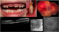 | 512 × 287 (250 KB) | Ozzie10aaaa | Author:Claire E L Smith, James A Poulter, Alex V Levin, Jenina E Capasso, Susan Price, Tamar Ben-Yosef, Reuven Sharony, William G Newman, Roger C Shore, Steven J Brookes, Alan J Mighell, and Chris F Inglehearn Leeds Institute of Biomedical and Clinical Sciences, St. James's University Hospital, University of Leeds,Sidney Kimmel Medical College at Thomas Jefferson University,Children's Hospital of the King's Daughters, Pediatric Ophthalmology and Ocular Genetics, Department of Clinical Genetic... |
File usage
The following page uses this file: