File:PMC4960283 11886 2016 761 Fig2 HTML (1).png

From WikiProjectMed
Jump to navigation Jump to search

PMC4960283_11886_2016_761_Fig2_HTML_(1).png(238 × 217 pixels, file size: 26 KB, MIME type: image/png)

This file is from a shared repository and may be used by other projects. The description on its file description page there is shown below.

Summary

Description
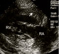
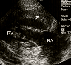
English: Fig2: A 20-year-old Hispanic G1P0 was transferred with a diagnosis of primary pulmonary hypertension at 27-week gestation. There was evidence of polycythemia (Hgb 16 g, Hct 48 %), finger clubbing, and central cyanosis. Pulmonary artery pressure was 90 mm Hg and reduced to 80 mm Hg with inhaled nitric oxide. a Parasternal long-axis view showing severe right ventricular enlargement (white line), a small left ventricular cavity, and paradoxical IVS motion. b RV inflow view showing marked right ventricular hypertrophy (white arrow). LV left ventricle, RA right atrium, RV right ventricle
Date
Source https://openi.nlm.nih.gov/detailedresult?img=PMC4960283_11886_2016_761_Fig2_HTML&query=Right%20ventricular%20hypertrophy&it=xg&req=4&npos=1
Author Narayanan M, Elkayam U, Naqvi TZ

Licensing

{{subst:Custom license marker added by UW}} https://creativecommons.org/licenses/by/4.0/ Attribution 4.0 International (CC BY 4.0)

This file was uploaded with UploadWizard.

File history

Click on a date/time to view the file as it appeared at that time.

Date/TimeThumbnailDimensionsUserComment
current21:45, 21 October 2022Thumbnail for version as of 21:45, 21 October 2022238 × 217 (26 KB)Ozzie10aaaaUploaded a work by Narayanan M, Elkayam U, Naqvi TZ from https://openi.nlm.nih.gov/detailedresult?img=PMC4960283_11886_2016_761_Fig2_HTML&query=Right%20ventricular%20hypertrophy&it=xg&req=4&npos=1 with UploadWizard

There are no pages that use this file.