File:PMC4897758 f1000research-5-9396-g0000.png
Jump to navigation
Jump to search
No higher resolution available.
PMC4897758_f1000research-5-9396-g0000.png (512 × 506 pixels, file size: 205 KB, MIME type: image/png)
File history
Click on a date/time to view the file as it appeared at that time.
| Date/Time | Thumbnail | Dimensions | User | Comment | |
|---|---|---|---|---|---|
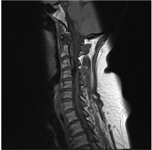
| current | 22:46, 26 August 2021 | | 512 × 506 (205 KB) | Ozzie10aaaa | Author:Iqbal Z, Mead P, Sayer JA Renal Services, Newcastle upon Tyne Hospitals NHS Foundation Trust (Openi/National Library of Medicine) Source:https://openi.nlm.nih.gov/detailedresult?img=PMC4897758_f1000research-5-9396-g0000&query=Gitelman%20syndrome&it=xg&req=4&npos=4 Description:f1: CT Cervical spine demonstrating a large ossified bony bar extending from the posterior surface of the C4 vertebral body up to the level of the upper surface of the C3 vertebral body.There are also multiple are... |
File usage
The following page uses this file: