File:PMC4661251 ceo-8-359-g001.png
Jump to navigation
Jump to search
No higher resolution available.
PMC4661251_ceo-8-359-g001.png (512 × 184 pixels, file size: 231 KB, MIME type: image/png)
File history
Click on a date/time to view the file as it appeared at that time.
| Date/Time | Thumbnail | Dimensions | User | Comment | |
|---|---|---|---|---|---|
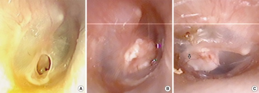
| current | 16:50, 1 November 2021 | 512 × 184 (231 KB) | Ozzie10aaaa | Author:Wu PW, Wang WH, Huang CC, Lee TJ, Huang CC ,Department of Otolaryngology-Head and Neck Surgery, Chang Gung Memorial Hospital (Openi/National Library of Medicine) Source:https://openi.nlm.nih.gov/detailedresult?img=PMC4661251_ceo-8-359-g001&query=perforated%20eardrum&it=xg&req=4&npos=13 Description:F1: Otoscope image of eardrum preoperatively (A), one month (B), and three months (C) postoperatively. Arrow indicates neovessel ingrowth on the outer surface of cartilage. |
File usage
The following page uses this file: