File:PMC4596109 jcn-11-372-g001 (1).png

From WikiProjectMed
Jump to navigation Jump to search

PMC4596109_jcn-11-372-g001_(1).png(512 × 566 pixels, file size: 747 KB, MIME type: image/png)

This file is from a shared repository and may be used by other projects. The description on its file description page there is shown below.

Summary

Description
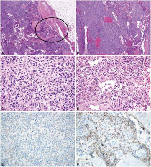
English: F1: Pathologic findings of case 2. Focal thymic carcinoma with an abundant thymoma (thymoma type B3) component. Low-power view showing a multinodular mass with extension to the peritumoral fat tissue [circle; hematoxylin-eosin stain (H-E); magnification, ×12] (A). The tumor is composed of abundant type B3 tissue (B: H-E; magnification, ×40; C: H-E, magnification, ×400) (B, C) and a focal thymic carcinoma component (squamous cell carcinoma) in the circled area (H-E; magnification, ×400) (D). Strong immunoreactivity for p63, a squamous cell differentiation marker, is found in the thymic carcinoma area (p63 immunostaining; magnification, ×400) (E). There is no p63 reactivity in the conventional thymoma B3 area (p63 immunostaining; magnification, ×400) (F).
Date
Source https://openi.nlm.nih.gov/detailedresult?img=PMC4596109_jcn-11-372-g001&query=Thymic%20carcinoma&it=xg&req=4&npos=2
Author Kim SH, Koh IS, Minn YK

Licensing

English: This file is licensed CC BY-NC 3.0

This file was uploaded with UploadWizard.

File history

Click on a date/time to view the file as it appeared at that time.

Date/TimeThumbnailDimensionsUserComment
current22:03, 11 November 2022Thumbnail for version as of 22:03, 11 November 2022512 × 566 (747 KB)Ozzie10aaaaUploaded a work by Kim SH, Koh IS, Minn YK from https://openi.nlm.nih.gov/detailedresult?img=PMC4596109_jcn-11-372-g001&query=Thymic%20carcinoma&it=xg&req=4&npos=2 with UploadWizard

The following page uses this file: