File:PMC4543185 kjped-58-256-g002.png
Jump to navigation
Jump to search
No higher resolution available.
PMC4543185_kjped-58-256-g002.png (512 × 208 pixels, file size: 102 KB, MIME type: image/png)
File history
Click on a date/time to view the file as it appeared at that time.
| Date/Time | Thumbnail | Dimensions | User | Comment | |
|---|---|---|---|---|---|
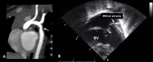
| current | 15:40, 20 October 2021 | 512 × 208 (102 KB) | Ozzie10aaaa | Author:Yoon JK, Ahn KJ, Kwon BS, Kim GB, Bae EJ, Noh CI, Ko JM, Department of Pediatrics, Seoul National University Children's Hospital, Seoul National University College of Medicine (Openi/National Library of Medicine) Source:https://openi.nlm.nih.gov/detailedresult?img=PMC4543185_kjped-58-256-g002&query=Kabuki%20syndrome&it=xg&req=4&npos=9 Description:F2: Congenital cardiac defects in Kabuki syndrome: left-sided heart anomalies. (A) Cardiac computed tomography reveals coarctation of the ao... |
File usage
The following page uses this file: