
No higher resolution available.
This file is from a shared repository and may be used by other projects.
The description on its file description page there is shown below.
License
Attribution 4.0 International (CC BY 4.0)
Summary
Author:Zong L, Guan J, Ealy M, Zhang Q, Wang D, Wang H, Zhao Y, Shen Z, Campbell CA, Wang F, Yang J, Sun W, Lan L, Ding D, Xie L, Qi Y, Lou X, Huang X, Shi Q, Chang S, Xiong W, Yin Z, Yu N, Zhao H, Wang J, Wang J, Salvi RJ, Petit C, Smith RJ, Wang Q ,Department of Otolaryngology-Head and Neck Surgery, Institute of Otolaryngology, PLA General Hospital(Openi/National Library of Medicine) Source:https://openi.nlm.nih.gov/detailedresult?img=PMC4518735_jmedgenet-2014-102961f04&query=Auditory%20neuropathy%20spectrum%20disorder&it=xg&req=4&npos=4 Description:JMEDGENET2014102961F4: Brain MRI imaging and muscle biopsy immune-staining of the patient (III: 3) from family 0223. (A) Serial cerebral MRI with fluid-attenuated inversion recovery sequence demonstrates normal signal intensity in bilateral centrum semiovale (left panel) and periventricular and subcortical white matter (right panel). (B) Axial view of the cerebellopontine angle and the internal auditory canal (IAC) shows normal anatomy (left panel). The two white lines illustrate the plane prescribed for oblique plane sagittal images obtained perpendicular to the nerves of the IAC. The oblique plane sagittal image (3D-fast-spin echo sequence, middle panel) obtained on the left side demonstrates an abnormally small cochlea nerve (Cn, white arrow) but a normal size IAC with normal facial (Fn), superior (Vsn) and inferior (Vin) vestibular nerves (green arrows). The right Cn was symmetrically small (right panel, white arrow). (C–F) Immunohistochemical staining of muscle biopsy (left gastrocnemius) in patient III: 3 shows a few atrophic myofibers (H&E, C). No ragged red fibres (modified Gomori-trichrome, D), ragged blue fibres (succinate dehydrogenase, E) or targetoid fibres (nicotinamide adenine dinucleotide-tetrazolium reductase, F) are identified. There is no reduction or absence of cytochrome-c-oxidase histochemical reactions observed (G).
File history
Click on a date/time to view the file as it appeared at that time.
| Date/Time | Thumbnail | Dimensions | User | Comment |
|---|
| current | 15:03, 18 November 2021 | 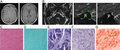 | 512 × 213 (186 KB) | Ozzie10aaaa | Author:Zong L, Guan J, Ealy M, Zhang Q, Wang D, Wang H, Zhao Y, Shen Z, Campbell CA, Wang F, Yang J, Sun W, Lan L, Ding D, Xie L, Qi Y, Lou X, Huang X, Shi Q, Chang S, Xiong W, Yin Z, Yu N, Zhao H, Wang J, Wang J, Salvi RJ, Petit C, Smith RJ, Wang Q ,Department of Otolaryngology-Head and Neck Surgery, Institute of Otolaryngology, PLA General Hospital(Openi/National Library of Medicine) Source:https://openi.nlm.nih.gov/detailedresult?img=PMC4518735_jmedgenet-2014-102961f04&query=Auditory%20neu... |
File usage
There are no pages that use this file.