File:PMC4463555 IJO-63-318-g002.png
Jump to navigation
Jump to search
No higher resolution available.
PMC4463555_IJO-63-318-g002.png (512 × 370 pixels, file size: 218 KB, MIME type: image/png)
File history
Click on a date/time to view the file as it appeared at that time.
| Date/Time | Thumbnail | Dimensions | User | Comment | |
|---|---|---|---|---|---|
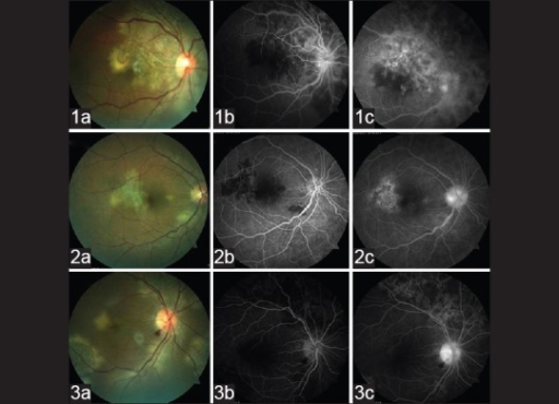
| current | 01:18, 1 February 2022 | | 512 × 370 (218 KB) | Ozzie10aaaa | Author:Venkatesh P, Gogia V, Gupta S, Tayade A, Shilpy N, Shah BM, Guleria R,Department of Ophthalmology, Dr. Rajendra Prasad Centre for Ophthalmic Sciences, All India Institute of Medical Sciences(Openi/National Library of Medicine) Source:https://openi.nlm.nih.gov/detailedresult?img=PMC4463555_IJO-63-318-g002&query=&req=4 Description:F1: Baseline fundus photographs of three representative patients (1a-3a) with fovea involving/threatening serpiginous choroiditis. Early phase of fluorescein a... |
File usage
There are no pages that use this file.