No higher resolution available.
This file is from a shared repository and may be used by other projects.
The description on its file description page there is shown below.
License
Attribution 3.0 Unported (CC BY 3.0)
Summary
Author:Fujinami K, Zernant J, Chana RK, Wright GA, Tsunoda K, Ozawa Y, Tsubota K, Robson AG, Holder GE, Allikmets R, Michaelides M, Moore AT, Laboratory of Visual Physiology, National Institute of Sensory Organs, National Hospital Organization, Tokyo Medical Center, Department of Ophthalmology, Keio University, School of Medicine, UCL Institute of Ophthalmology, Moorfields Eye Hospital (Openi/National Library of Medicine) Source:https://openi.nlm.nih.gov/detailedresult?img=PMC4459618_nihms693250f3&query=Stargardt%20disease&it=xg&req=4&npos=12
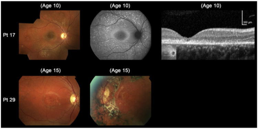
Description:F3: Color fundus photographs, autofluorescence (AF), and spectral-domain optical coherence tomographic images of 2 molecularly proven cases with “atypical” clinical features of childhood-onset Stargardt Disease (patients 17 and 29). Color photograph of patient 17 shows fine dots at the central macula surrounded by numerous peripheral flecks and AF imaging demonstrates well-defined dots associated with a high signal at the central macula surrounded by a ring of increased AF signal and numerous foci with high and low signal extending to the peripheral retina. Outer retinal loss at the macula is present on SD-OCT. Patient 29 has asymmetric fundus findings with central atrophy and peripheral flecks in the right eye and macular atrophy with flecks, subretinal fibrosis, and hyperpigmentation at the level of the retinal pigment epithelium in the left eye. Pt = patient.
File history
Click on a date/time to view the file as it appeared at that time.
| Date/Time | Thumbnail | Dimensions | User | Comment |
|---|
| current | 15:01, 25 August 2021 |  | 512 × 256 (134 KB) | Ozzie10aaaa | Author:Fujinami K, Zernant J, Chana RK, Wright GA, Tsunoda K, Ozawa Y, Tsubota K, Robson AG, Holder GE, Allikmets R, Michaelides M, Moore AT, Laboratory of Visual Physiology, National Institute of Sensory Organs, National Hospital Organization, Tokyo Medical Center, Department of Ophthalmology, Keio University, School of Medicine, UCL Institute of Ophthalmology, Moorfields Eye Hospital (Openi/National Library of Medicine) Source:https://openi.nlm.nih.gov/detailedresult?img=PMC4459618_nihms... |
File usage
The following page uses this file: