File:PMC4322316 BMRI2015-545243.002.png
Jump to navigation
Jump to search
No higher resolution available.
PMC4322316_BMRI2015-545243.002.png (512 × 277 pixels, file size: 143 KB, MIME type: image/png)
File history
Click on a date/time to view the file as it appeared at that time.
| Date/Time | Thumbnail | Dimensions | User | Comment | |
|---|---|---|---|---|---|
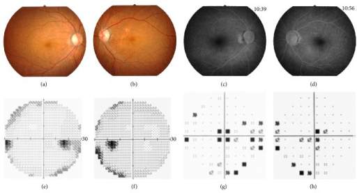
| current | 22:18, 29 October 2021 | | 512 × 277 (143 KB) | Ozzie10aaaa | Author:Kikuchi S, Kameya S, Gocho K, El Shamieh S, Akeo K, Sugawara Y, Yamaki K, Zeitz C, Audo I, Takahashi H,Department of Ophthalmology, Nippon Medical School, Chiba Hokuso Hospital (Openi/National Library of Medicine) Source:https://openi.nlm.nih.gov/detailedresult?img=PMC4322316_BMRI2015-545243.002&query=&req=4 Description:fig2: Clinical findings of the proband, Patient II-2. Fundus photographs (a, b) and fluorescein angiograms (c, d) show no abnormal findings. The results of the Humphrey... |
File usage
The following page uses this file: