File:PMC4273719 parasite-21-74-fig1.png
Jump to navigation
Jump to search
No higher resolution available.
PMC4273719_parasite-21-74-fig1.png (512 × 188 pixels, file size: 80 KB, MIME type: image/png)
File history
Click on a date/time to view the file as it appeared at that time.
| Date/Time | Thumbnail | Dimensions | User | Comment | |
|---|---|---|---|---|---|
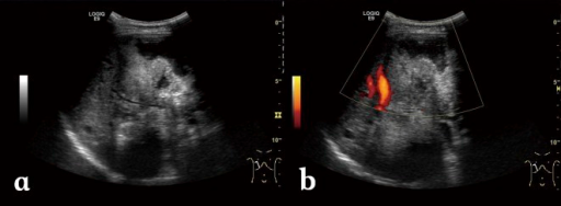
| current | 21:15, 15 November 2021 | 512 × 188 (80 KB) | Ozzie10aaaa | Author:Liu W, Delabrousse É, Blagosklonov O, Wang J, Zeng H, Jiang Y, Wang J, Qin Y, Vuitton DA, Wen H,Imaging Center, First Affiliated Hospital, Xinjiang Medical University Hospital(Openi/National Library of Medicine) Source:https://openi.nlm.nih.gov/detailedresult?img=PMC4273719_parasite-21-74-fig1&query=Alveolar%20echinococcosis&it=xg&req=4&npos=17 Description:F1: Alveolar echinococcosis in a 30-year-old woman. (a) Abdominal gray-scale US image shows an irregular type heterogeneous mass le... |
File usage
There are no pages that use this file.