File:PMC4198596 ad-26-639-g001.png
Jump to navigation
Jump to search
No higher resolution available.
PMC4198596_ad-26-639-g001.png (512 × 379 pixels, file size: 288 KB, MIME type: image/png)
File history
Click on a date/time to view the file as it appeared at that time.
| Date/Time | Thumbnail | Dimensions | User | Comment | |
|---|---|---|---|---|---|
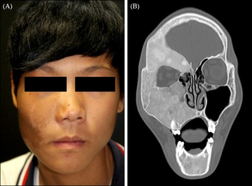
| current | 20:45, 7 August 2021 | | 512 × 379 (288 KB) | Ozzie10aaaa | Author:Jung KE, Lee JH, Kim TY,Department of Dermatology, Eulji University Hospital, Daejeon, Korea (Openi/National Library of Medicine) Source:https://openi.nlm.nih.gov/detailedresult?img=PMC4198596_ad-26-639-g001&query=Polyostotic%20fibrous%20dysplasia&it=xg&req=4&npos=25 Description:F1: Solitary, brown patch with irregular border, throughout the patient's right cheek and upper eyelid with facial asymmetry. (B) Coronal computed tomography scan demonstrates heterogenous ground glass appearan... |
File usage
The following page uses this file: