File:PMC4131998 dp0403a08g003.png
Jump to navigation
Jump to search
No higher resolution available.
PMC4131998_dp0403a08g003.png (512 × 379 pixels, file size: 450 KB, MIME type: image/png)
File history
Click on a date/time to view the file as it appeared at that time.
| Date/Time | Thumbnail | Dimensions | User | Comment | |
|---|---|---|---|---|---|
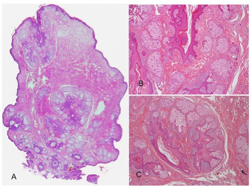
| current | 21:06, 10 March 2022 | | 512 × 379 (450 KB) | Ozzie10aaaa | Author:Watanabe-Okada E, Kurihara Y, Miyakawa S, Tanaka M,Division of Dermatology, Kawasaki Municipal Hospital(Openi/National Library of Medicine)Source:https://openi.nlm.nih.gov/detailedresult?img=PMC4131998_dp0403a08g003&query=&req=4 Description:f3-dp0403a08: (A) Histopathologic examination reveals an exophytic lesion consisting of a dilated infundibular cystic structure with sebaceous lobules connected via sebaceous ducts. (H&E ×5). (B) Numerous mature sebaceous lobules along with the elon... |
File usage
The following page uses this file: