File:PMC4105805 kjr-15-439-g001.png
Jump to navigation
Jump to search
No higher resolution available.
PMC4105805_kjr-15-439-g001.png (512 × 263 pixels, file size: 217 KB, MIME type: image/png)
File history
Click on a date/time to view the file as it appeared at that time.
| Date/Time | Thumbnail | Dimensions | User | Comment | |
|---|---|---|---|---|---|
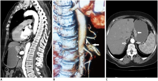
| current | 23:30, 16 January 2022 | | 512 × 263 (217 KB) | Ozzie10aaaa | Author:Gunduz Y, Asil K, Aksoy YE, Tatlı Ayhan L,Department of Radiology, Sakarya University Medical Faculty(Openi/National Library of Medicine)Source:https://openi.nlm.nih.gov/detailedresult?img=PMC4105805_kjr-15-439-g001&query=Median%20arcuate%20ligament%20syndrome&it=xg&req=4&npos=1 Description:F1: Median arcuate ligament syndrome in 72-year-old male patient.A. Sagittal reformatted contrast enhanced CT angiography shows stenosis and aneurysm of celiac artery due to compression by median ar... |
File usage
There are no pages that use this file.