No higher resolution available.
This file is from a shared repository and may be used by other projects.
The description on its file description page there is shown below.
License
Attribution 3.0 Unported (CC BY 3.0)
Summary
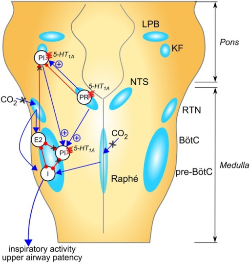
Author:Abdala AP, Bissonnette JM, Newman-Tancredi A ,School of Physiology and Pharmacology, University of Bristol (Openi/National Library of Medicine) Source:https://openi.nlm.nih.gov/detailedresult?img=PMC4038922_fphys-05-00205-g0001&query=Rett%20syndrome&it=xg&req=4&npos=1 Description:F1: Suggested mechanisms for respiratory rhythm disease in Rett syndrome and network targets of 5-HT1A agonists. Populations of respiratory neurons are shown in white circles (see below). Blue arrows indicate excitatory drive; red connectors with circle-ends indicate inhibitory drive. A healthy respiratory rhythm and pattern are critically dependent on the balance between excitatory and inhibitory synaptic drives to the “core” of mutually inhibitory respiratory neurons located in the BötC and pre-BötC (Smith et al., 2013). The disturbed rhythm in Rett syndrome seems to arise from an imbalance of drives to this core circuitry (indicated by black “X” when reduced, or blue “+” when enhanced); many mechanisms contribute to this: (i) weakened excitatory synaptic drives to and within the inspiratory “kernel” (Viemari et al., 2005), (ii) reduced CO2 sensitivity (Zhang et al., 2011; Toward et al., 2013; Bissonnette et al., 2014); (iii) excess descending post-inspiratory drive from the pontine parabrachial complex (Stettner et al., 2007; Voituron et al., 2010; Dhingra et al., 2013); which could be a consequence of loss of inhibitory drives to this area, including KF (Stettner et al., 2007; Abdala et al., 2010). In combination, these mechanisms would lead to disinhibition of PI populations, disruption of timing for termination of inspiration and expiratory length irregularity. Studies in humans and mice suggest that breath-holds and Valsalva maneuvers may be linked to active closure of the glottis implicating a failure in the ponto-medullary gating of central post-inspiratory activity, for a review; see Ramirez et al. (2013). 5-HT1A receptors suppress specific inhibitory glycinergic neuron populations in the “core” of mutually inhibitory neurons with consequent disinhibition of inspiratory populations (Shevtsova et al., 2011). In addition, 5-HT1A receptors can directly reduce the activity of neuron populations contributing to the descending post-inspiratory drive from the pons. I, inspiratory neuron population; PI, post-inspiratory neuron population; E2, late expiratory neuron population; PR, pulmonary stretch relay; LBP, lateral parabrachial nu; KF, Kölliker-Fuse nu; NTS, nucleus of the solitary tract; RTN, retrotrapezoid nu; BötC, Bötzinger complex; pre-BötC, pre-Bötzinger complex.
File history
Click on a date/time to view the file as it appeared at that time.
| Date/Time | Thumbnail | Dimensions | User | Comment |
|---|
| current | 20:03, 13 October 2021 |  | 512 × 537 (267 KB) | Ozzie10aaaa | Author:Abdala AP, Bissonnette JM, Newman-Tancredi A ,School of Physiology and Pharmacology, University of Bristol (Openi/National Library of Medicine) Source:https://openi.nlm.nih.gov/detailedresult?img=PMC4038922_fphys-05-00205-g0001&query=Rett%20syndrome&it=xg&req=4&npos=1 Description:F1: Suggested mechanisms for respiratory rhythm disease in Rett syndrome and network targets of 5-HT1A agonists. Populations of respiratory neurons are shown in white circles (see below). Blue arrows indicate... |
File usage
The following page uses this file: