File:PMC4017419 GHFBB-4-164-g001.png
Jump to navigation
Jump to search

Size of this preview: 429 × 600 pixels. Other resolutions: 171 × 240 pixels | 512 × 716 pixels.
Original file (512 × 716 pixels, file size: 940 KB, MIME type: image/png)
File history
Click on a date/time to view the file as it appeared at that time.
| Date/Time | Thumbnail | Dimensions | User | Comment | |
|---|---|---|---|---|---|
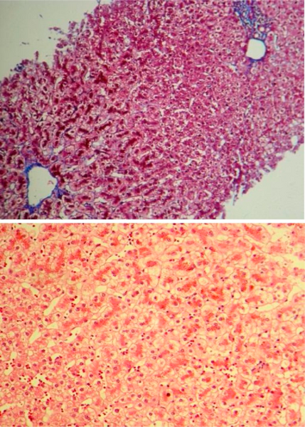
| current | 15:43, 12 October 2021 | | 512 × 716 (940 KB) | Ozzie10aaaa | Author:Lahmi F, Roshani M, Khosravi K, Azizi M, Mohebbi SR, Zali MR ,Research Institute for Gastroenterology and Liver Disease, Shahid Beheshti University of Medical sciences (Openi/National Library of Medicine) Source:https://openi.nlm.nih.gov/detailedresult?img=PMC4017419_GHFBB-4-164-g001&query=Dubin%E2%80%93Johnson%20syndrome&it=xg&req=4&npos=2 Description:F0001: Histopathological view of liver needle biopsy shows intact lobular and vascular architecture. Individual hepatocytes contain abu... |
File usage
The following page uses this file: