No higher resolution available.
This file is from a shared repository and may be used by other projects.
The description on its file description page there is shown below.
License
Attribution-NonCommercial-NoDerivs 3.0 Unported (CC BY-NC-ND 3.0)
Summary
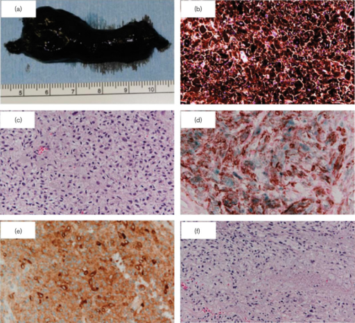
Author:Nitta K, Kashima T, Mayuzumi H, Akiyama H, Miyanaga T, Hirato J, Kishi S,Department of Ophthalmology, Gunma University School of Medicine,Clinical Department of Pathology, Gunma University Hospital(Open/National Library of Medicine) Source:https://openi.nlm.nih.gov/detailedresult?img=PMC4004635_cmr-24-286-g002&query=Animal-type%20melanoma&it=xg&req=4&npos=4 Description:F2: (a) Macroscopy of the extracted orbital tumor: highly pigmented 6×2 cm large tumor, which ruptured during the extirpation. (b) Hematoxylin and eosin staining of the orbital tumor (×20): melanin-containing cells (melanocytes and melanophages) are abundant in the tumor. (c) Hematoxylin and eosin staining of the orbital tumor after using a bleaching method for melanins (×20): diffuse proliferation of the epithelioid cells and spindle cells, and low-grade nuclear atypia were seen. Small nucleoli were seen in some areas of the tissue. (d) HMB (Human Melanoma Black)-45 staining (×20): positive for atypical cells. (e) Melan-A staining (×20): positive for atypical cells. (f) Hematoxylin and eosin staining of the orbital tumor after using a bleaching method for melanins (×20): vast areas of necrosis were seen.
File history
Click on a date/time to view the file as it appeared at that time.
| Date/Time | Thumbnail | Dimensions | User | Comment |
|---|
| current | 21:38, 29 March 2022 |  | 512 × 466 (566 KB) | Ozzie10aaaa | Author:Nitta K, Kashima T, Mayuzumi H, Akiyama H, Miyanaga T, Hirato J, Kishi S,Department of Ophthalmology, Gunma University School of Medicine,Clinical Department of Pathology, Gunma University Hospital(Open/National Library of Medicine) Source:https://openi.nlm.nih.gov/detailedresult?img=PMC4004635_cmr-24-286-g002&query=Animal-type%20melanoma&it=xg&req=4&npos=4 Description:F2: (a) Macroscopy of the extracted orbital tumor: highly pigmented 6×2 cm large tumor, which ruptured during the exti... |
File usage
There are no pages that use this file.
.png)
.png)