File:PMC3952380 JCIS-4-5-g003.png
Jump to navigation
Jump to search
No higher resolution available.
PMC3952380_JCIS-4-5-g003.png (512 × 207 pixels, file size: 237 KB, MIME type: image/png)
File history
Click on a date/time to view the file as it appeared at that time.
| Date/Time | Thumbnail | Dimensions | User | Comment | |
|---|---|---|---|---|---|
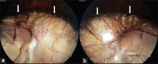
| current | 18:32, 23 October 2021 | 512 × 207 (237 KB) | Ozzie10aaaa | Author:Natung T, Goyal A, Handique A, Kapoor M ,Department of Ophthalmology, North Eastern Indira Gandhi Regional Institute of Health and Medical Sciences (Openi/National Library of Medicine) Source:https://openi.nlm.nih.gov/detailedresult?img=PMC3952380_JCIS-4-5-g003&query=CHARGE%20SYNDROME&it=xg&req=4&npos=11 Description:F2: 27-year-old female with poor vision and inability to hear was subsequently diagnosed with CHARGE syndrome. (a and b) Fundus photographs (antero-posterior view) show bil... |
File usage
The following page uses this file: