File:PMC3895851 1477-7819-12-8-2.png
Jump to navigation
Jump to search
No higher resolution available.
PMC3895851_1477-7819-12-8-2.png (512 × 466 pixels, file size: 185 KB, MIME type: image/png)
File history
Click on a date/time to view the file as it appeared at that time.
| Date/Time | Thumbnail | Dimensions | User | Comment | |
|---|---|---|---|---|---|
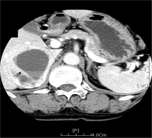
| current | 00:17, 21 February 2022 | | 512 × 466 (185 KB) | Ozzie10aaaa | Author:Tang KL, Lin Y, Li LM, Department of Urology, General Hospital of Tianjin Medical University(Openi/National Library of medicine) Source:https://openi.nlm.nih.gov/detailedresult?img=PMC3895851_1477-7819-12-8-2&query=&req=4 Description:F2: Enhanced CT scanning of the adrenal gland shows a round-like cystic solid mass located at the right adrenal gland (Black arrow). Inside of the mass, multiple punctate and dense shadows are visible. The boundary of the mass is manifested as inhomogeneou... |
File usage
The following page uses this file: