File:PMC3849310 kjo-27-454-g001.png
Jump to navigation
Jump to search
No higher resolution available.
PMC3849310_kjo-27-454-g001.png (512 × 388 pixels, file size: 335 KB, MIME type: image/png)
File history
Click on a date/time to view the file as it appeared at that time.
| Date/Time | Thumbnail | Dimensions | User | Comment | |
|---|---|---|---|---|---|
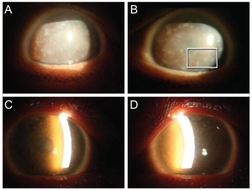
| current | 18:47, 12 August 2021 | | 512 × 388 (335 KB) | Ozzie10aaaa | Author:Lee YK, Chang DJ, Chung SK.Department of Ophthalmology, The Catholic University of Korea College of Medicine (Openi/National Library of Medicine) Source:https://openi.nlm.nih.gov/detailedresult?img=PMC3849310_kjo-27-454-g001&query=Macular%20corneal%20dystrophy&it=xg&req=4&npos=7 Description: F1: Slit lamp photography of the patient (A,B). (A) Right eye and (B) left eye. Diffusely hazy corneas with bilateral opacities were observed. There were multiple irregular, grayish-white, dense, p... |
File usage
The following page uses this file: