File:PMC3771223 ijms-38-191-g001.png
Jump to navigation
Jump to search
No higher resolution available.
PMC3771223_ijms-38-191-g001.png (512 × 168 pixels, file size: 194 KB, MIME type: image/png)
File history
Click on a date/time to view the file as it appeared at that time.
| Date/Time | Thumbnail | Dimensions | User | Comment | |
|---|---|---|---|---|---|
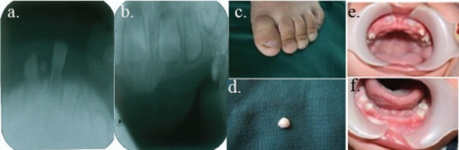
| current | 20:10, 13 January 2022 | 512 × 168 (194 KB) | Ozzie10aaaa | Author:Ghaderi F, Hekmat S, Ghaderi R, Fardaei M,Department of Pediatric Dentistry, School of Dentistry, Shiraz University of Medical Sciences (Openi/National library of Medicine) Source:https://openi.nlm.nih.gov/detailedresult?img=PMC3771223_ijms-38-191-g001&query=Witkop%20syndrome&it=xg&req=4&npos=1 Description:F1: These are the clinical and radiographic manifestations of the patient’s condition. a. The mandibular anterior permanent germ in periapical view. b. The maxillary anterior permane... |
File usage
There are no pages that use this file.