File:PMC3698900 IJRI-22-358-g003.png
Jump to navigation
Jump to search
No higher resolution available.
PMC3698900_IJRI-22-358-g003.png (512 × 189 pixels, file size: 265 KB, MIME type: image/png)
File history
Click on a date/time to view the file as it appeared at that time.
| Date/Time | Thumbnail | Dimensions | User | Comment | |
|---|---|---|---|---|---|
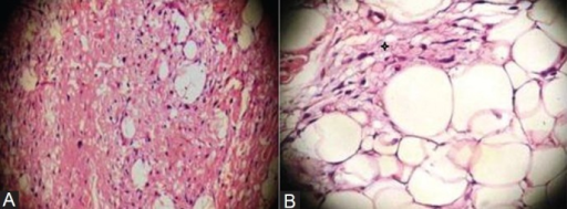
| current | 18:22, 19 April 2022 | 512 × 189 (265 KB) | Ozzie10aaaa | Author:Kumar N, Mittal M, Sinha M, Thukral B ,Department of Radio-Diagnosis, VM Medical College and Safdarjung Hospital (Openi/National of Medicine) Source:https://openi.nlm.nih.gov/detailedresult?img=PMC3698900_IJRI-22-358-g003&query=Neural%20fibrolipoma&it=xg&req=4&npos=4 Description:F3: Photomicrograph of the histopathology specimen (Haematoxylin and eosin; original and 200x magnification). The lesion is composed of uniform appearing adipocytes with strands of spindle cells (black star) |
File usage
There are no pages that use this file.